. 2025 Jan-Mar;66(1):179-197.
doi: 10.47162/RJME.66.1.17.

Immunoprofile of some surface and cytoplasmic peripheral cell adhesion molecules in oral squamous cell carcinoma

Affiliations

Immunoprofile of some surface and cytoplasmic peripheral cell adhesion molecules in oral squamous cell carcinoma

Ionuţ Octavian Ilie et al. Rom J Morphol Embryol. 2025 Jan-Mar.

Abstract

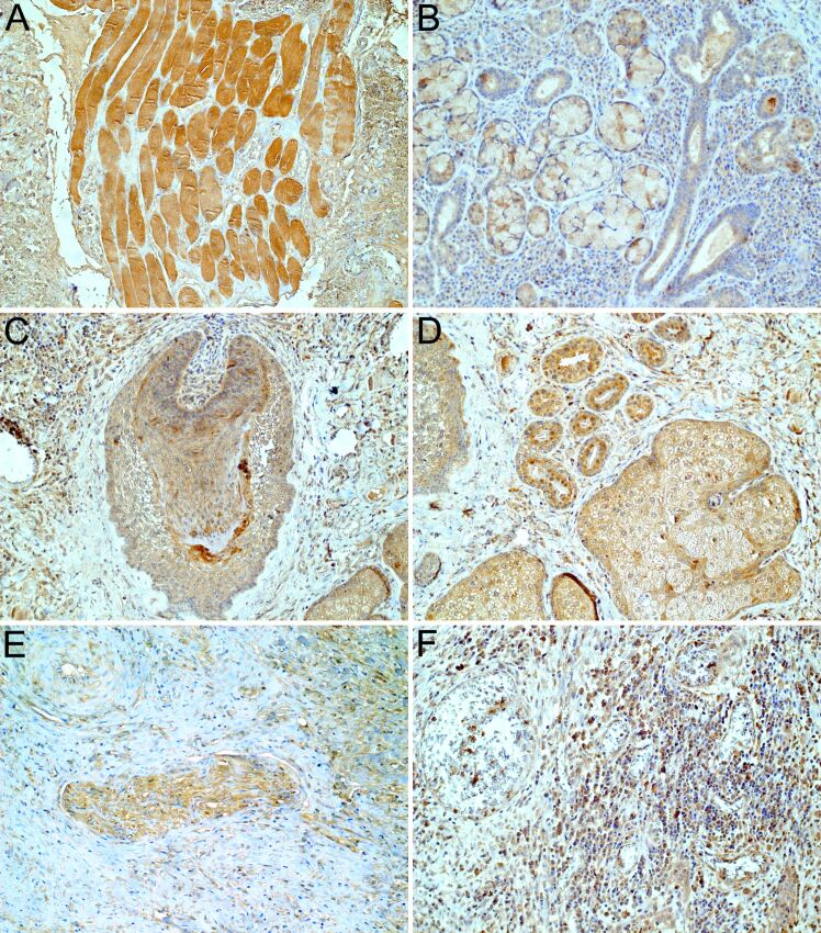
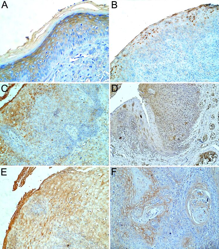
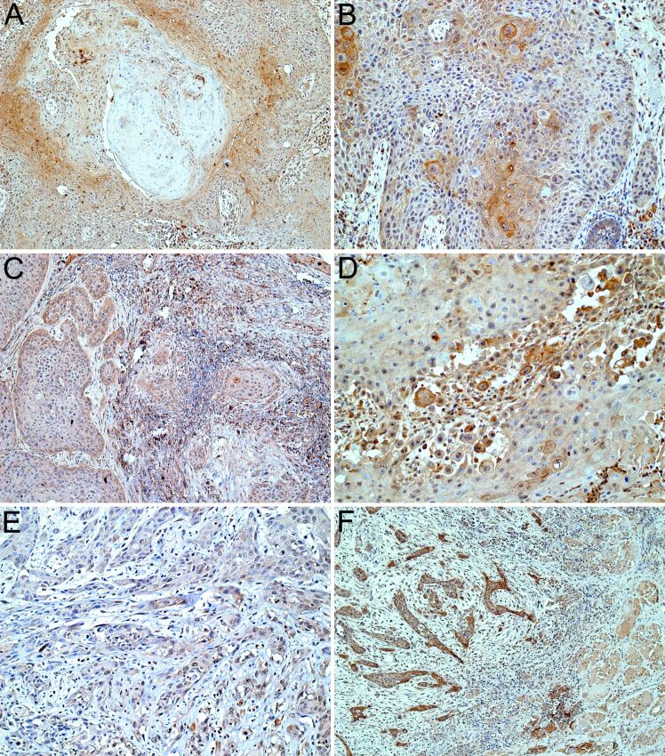
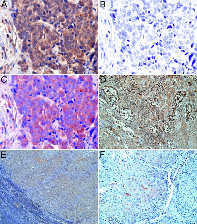
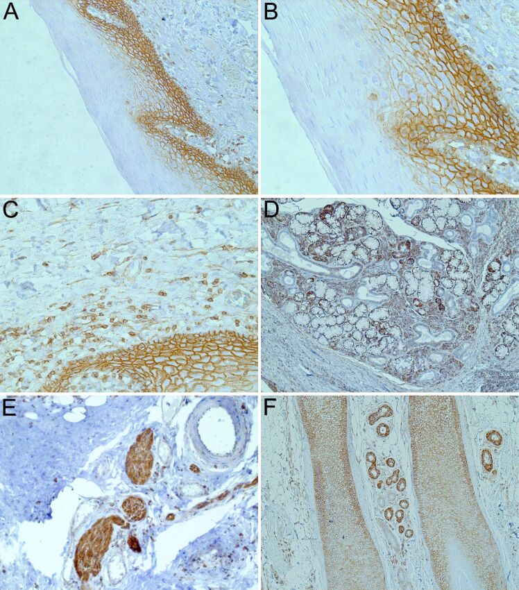
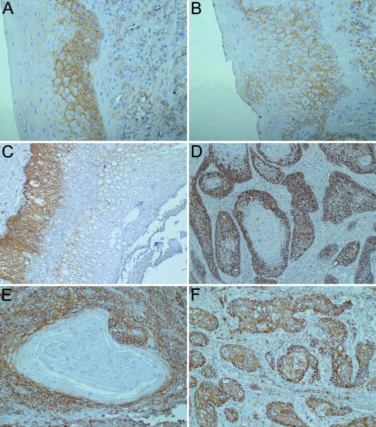
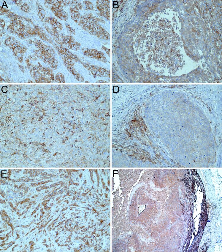
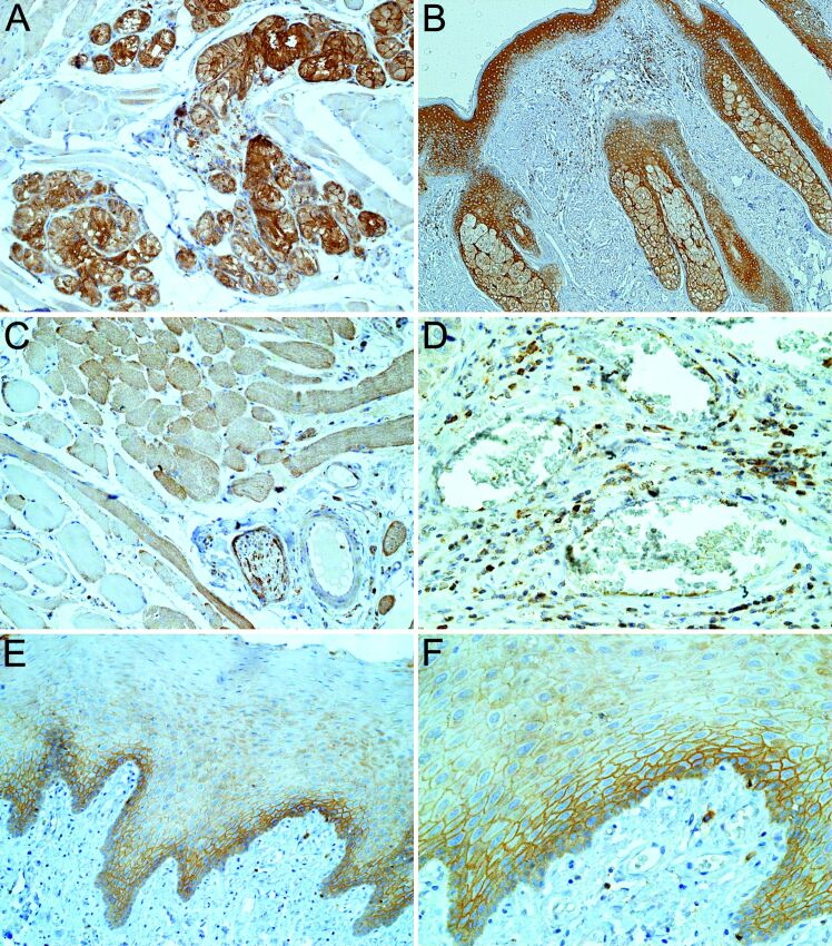
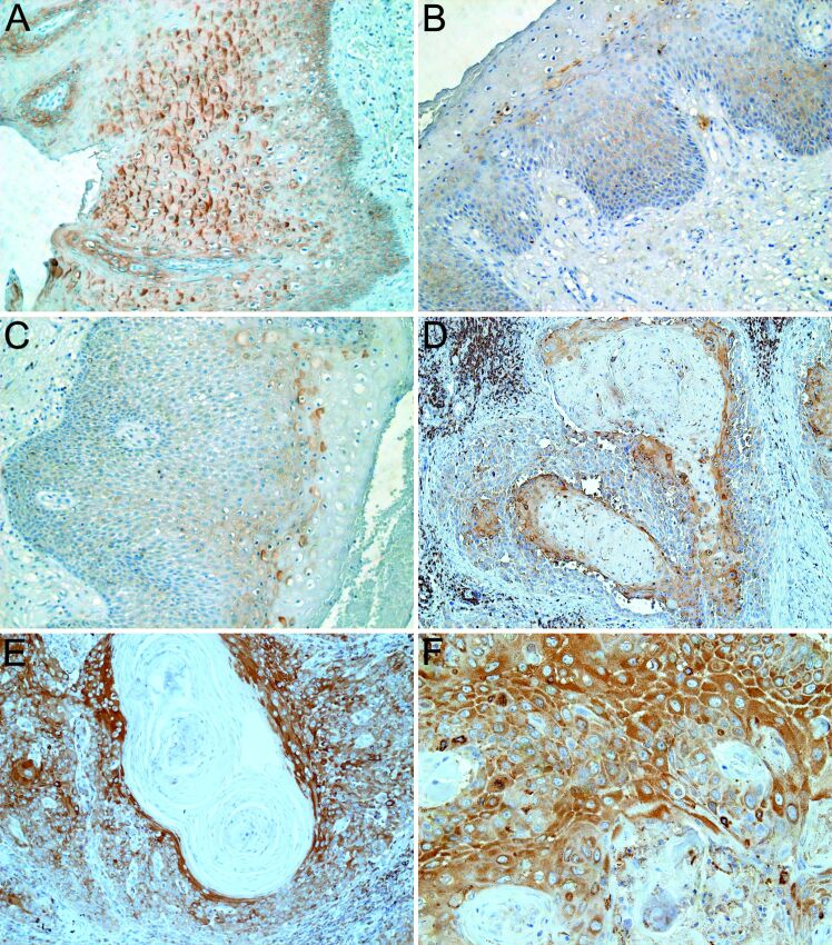
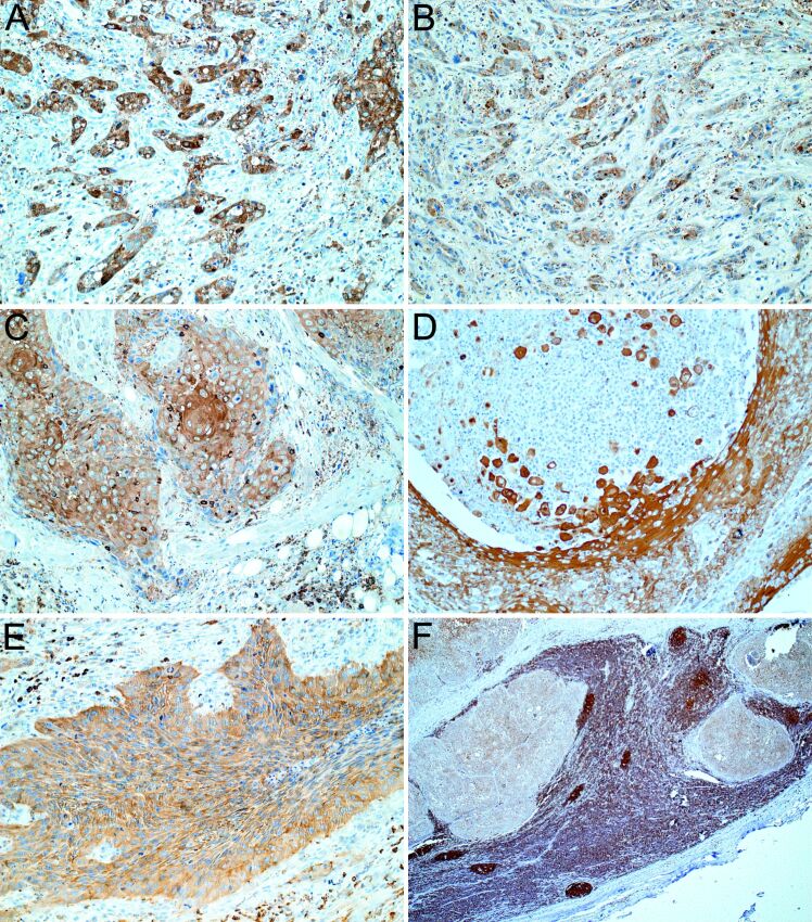
Despite the recent advances in diagnosis and treatment, oral squamous cell carcinoma (OSCC) continues to have a low overall survival rate (around 50%), being a tumor with high locoregional aggressiveness and high risk of lymph node (LN) dissemination. Such behavior can also be explained by the alteration of the expression of adhesion molecules, allowing tumor cells to invade surrounding tissues and make them capable of metastasizing. In this regard, we initiated a study on the immunohistochemical expression of Integrin alphavbeta6 (Integrin αvβ6), CD44 and Ezrin in OSCCs. A number of 39 such tumors with various locations in the oral cavity were investigated by enzymatic immunohistochemistry together with several samples of oral mucosa and oral dysplastic lesions. Using integrated optical density (IOD) as a method to quantify, we observed that the reactivity of these three markers decreased in the progression of dysplastic lesions and in the transition from well to moderately and poorly differentiated tumors. Also, in both conditions we noticed a shift of pattern reactivity from the continuous membrane to discontinuous membrane and cytoplasmic one, even to a nuclear pattern. In addition, the reactivity of the three markers was more evident in the invasion front and especially in these tumors with discohesive growth patterns. All this suggests the involvement of these adhesion molecules in the processes of transformation and malignant progression of OSCCs. It also explains their possible involvement in locoregional aggressiveness and LN dissemination.

Keywords: CD44; Ezrin; Integrin αvβ6; dysplastic lesions; immunohistochemistry; oral squamous cell carcinoma.


Conflict of interest statement

The authors declare that there is no conflict of interests regarding the publication of this paper. All authors read and approved the final manuscript.

Figures

Figure 1
Figure 1
Integrin αvβ6 immunoreactivity: (A) At the level of striated muscle fibers, where the cytoplasmic pattern predominated; (B) At the level of minor salivary glands, with cytoplasmic and membrane pattern, the reactivity being more evident in the serous acini; (C) At the level of hairs from the lips, with predominant cytoplasmic reactivity; (D) At the level of sebaceous and sweating glands from the lips, with predominant cytoplasmic and nuclear staining (the latter being more evident in the luminal cells of the sweat glands); (E) A weak reactivity observed at the level of the nerve threads; (F) At the level of inflammatory cells (especially in lymphocytes and plasma cells) and in the endothelium of blood vessels, with predominantly cytoplasmic reactivity. Anti-Integrin αvβ6 immunolabeling: (A–F) ×200
Figure 2
Figure 2
Integrin αvβ6 immunoreactivity: (A) At the level of normal epithelium, basal cells were not reactive or the cytoplasmic reactivity was weak, but the membrane pattern was especially present at the level of the upper rows of the spinous layer; (B) In low-grade dysplasia, we noticed the tendency to change the subcellular pattern from the membranous to the cytoplasmic to the upper rows of the spinous layer; (C) In moderate-grade dysplasia, the same trend was observed as in low-grade dysplasia, but in the inner two-thirds the reactivity was absent or present with a weak membranous pattern; (D) In high-grade dysplasia, we noticed a nuclear reactivity extended throughout the entire thickness of the epithelium and a weak cytoplasmic reactivity in the upper part of the epithelium; (E) In the epithelium with koilocytic atypia, the reactivity was present in the peripheral cytoplasm, being more evident in the upper layers; (F) In well-differentiated tumors, the prevailing pattern it was membranous especially inside the tumor islands at the level of neoplastic cells with morphology similar to normal keratinocytes. Anti-Integrin αvβ6 immunolabeling: (A–C, E and F) ×200; (D) ×100
Figure 3
Figure 3
Integrin αvβ6 immunoreactivity: (A) In well-differentiated OSCC, the ortho- and parakeratotic pearls were negative; (B) In the moderately differentiated OSCC, the predominant reactivity is cytoplasmic, maintaining membrane reactivity only at the level of well-differentiated neoplastic cells; (C) In the less differentiated OSCC, cells lose their membrane reactivity and acquire a predominantly cytoplasmic and even nuclear pattern more evident towards the periphery of the proliferations and in the invasion front; (D) An intense membrane and cytoplasmic reactivity observed at the level of acantholytic neoplastic cells in those variants with extensive foci of acantholysis; (E) In the poorly differentiated OSCC, pattern of reactivity is the same as in moderately differentiated variant with an intensification of the nuclear staining; (F) Regardless of the degree of differentiation, the reactivity was more evident in the invasion front, the invasive islands having a predominantly cytoplasmic and nuclear pattern. Anti-Integrin αvβ6 immunolabeling: (A, C and F) ×100; (B, D and E) ×200. OSCC: Oral squamous cell carcinoma
Figure 4
Figure 4
Integrin αvβ6 immunoreactivity: (A) Spectral library built within the Nuance FX package in order to evaluate the nuclear immunohistochemical expression of the Integrin αvβ6 – raw RGB exemplary image of Integrin αvβ6 immunohistochemistry, as captured by the sensor in RGB mode; (B and C) Correspondent unmixed images showing separate spectra for Hematoxylin, as well as the merged image of DAB and Hematoxylin, clearly show nuclear staining for Integrin αvβ6; (D) Intense reactivity at the level of the tumor stroma, especially in those cases with inflammatory stroma, the lymphocytes and vascular endothelial cells being intensely positive; (E and F) A predominantly weak cytoplasmic reactivity at the level of metastatic proliferations from the lymph nodes. Anti-Integrin αvβ6 immunolabeling: (A–C) ×400; (D and F) ×200; (E) ×100. DAB: 3,3’-Diaminobenzidine; RGB: Red, green and blue
Figure 5
Figure 5
CD44 immunoreactivity: (A) In the normal oral epithelial tissue, the reactivity was present in its lower half, predominantly with membrane pattern, the reactivity decreasing progressively from the basal layer to the spinous layer; (B) At the level of the basal cells, the reactivity was absent at the interface with the chorion, and in addition, in the cells of the basal and parabasal layers, CD44 was also weakly positive into the cytoplasm; (C) At the level of the chorion, CD44 was positive in inflammatory cells, especially in lymphocytes, in vascular endothelial cells and in fibroblasts; (D) A membrane and cytoplasmic reactivity was recorded in minor salivary gland lobules, especially at the level of serous acini; (E) Intense reactivity at the level of peripheral nerves; (F) In the lip specimens, we recorded CD44 reactivity with prevailing membranous pattern at the level of the hairs and sweat glands. Anti-CD44 immunolabeling: (A, D and E) ×200; (B and D) ×400; (F) ×100. CD44: Cluster of differentiation 44
Figure 6
Figure 6
CD44 immunoreactivity: (A) In low-grade dysplasia, the CD44 reactivity was present in the lower third of the epithelium especially in the parabasal layer and in the rows above it; (B) In moderate-grade dysplasia, the reactivity was lower than in the low grade, but still extended to close to the surface; (C) In cases associated with koilocytic atypia, a weak but discontinuous membrane reactivity was observed, especially at the level of koilocytes from the lower layers; (D) In the well-differentiated tumor, we recorded the highest reactivity, the pattern being membranous, continuous, more evident in the periphery of proliferations; (E) The reactivity was absent at the level of ortho- and parakeratotic pearls; (F) In moderately differentiated tumor, the continuous membranous pattern was more obvious at the periphery of tumor islands; inside them, the pattern was membranous but discontinuous and also diffuse cytoplasmic. Anti-CD44 immunolabeling: (A and B) ×400; (C, E and F) ×200; (D) ×100. CD44: Cluster of differentiation 44
Figure 7
Figure 7
CD44 immunoreactivity: (A) At the invasion front, sometimes the membrane reactivity of tumor proliferations was absent at the interface with the stroma; (B) In the acantholytic variant, the membrane reactivity is also present in the acantholytic cells, similar to that from the well-differentiated neoplastic cells with spinous cells morphology; (C) The poorly differentiated tumor presented lower reactivity, with a discontinuous membrane pattern and a cytoplasmic one; (D) In the basaloid variant, the reactivity was low and was present inside the tumor islands, with the dominant pattern as a cytoplasmic one; (E) The highest reactivity was recorded in tumors with discohesive growth pattern; (F) A weak reactivity, predominantly cytoplasmic, was also observed at the level of tumor proliferations from lymph node metastases. Anti-CD44 immunolabeling: (A–E) ×200; (F) ×50. CD44: Cluster of differentiation 44
Figure 8
Figure 8
Ezrin immunoreactivity: (A) Reactivity at level of minor salivary glands, more intense in glandular cells than in ductal cells with membranous and cytoplasmic pattern; (B) At the level of lip samples, we noticed an intends reaction in epidermis and skin appendages; (C) A weak reaction was observed at the level of striated muscle fibers and nerve filaments; (D) Reactivity at the level of blood endothelial cells and stromal inflammatory cells (lymphocytes); (E) At the level of oral epithelium, we noticed membranous expression in the parabasal layer and stratum spinosum, especially in the deep layers; (F) A weak membranous expression was also noticed at the lateral and apical surfaces of basal cells but not at the stromal interface. Anti-Ezrin immunolabeling: (A, C and E) ×200; (B) ×100; (D and F) ×400
Figure 9
Figure 9
Ezrin immunoreactivity: (A) In koilocytes was noticed an intensification of staining in the peripheral cytoplasm from their lower part; (B) In low-grade dysplastic lesions prevail the membrane reactivity; (C) In high-grade dysplastic lesions prevail the cytoplasm reactivity; (D) In well-differentiated tumors, keratin pearls were negative; (E) The most intense reaction was recorded in more differentiated keratinized cells with both membrane and cytoplasmic pattern, with the last one being much more pronounced; (F) In moderately differentiated tumors, the reactivity was much more obvious and with a predominant cytoplasmic pattern. Anti-Ezrin immunolabeling: (A–C and E) ×200; (D) ×100; (F) ×400
Figure 10
Figure 10
Ezrin immunoreactivity: (A and B) In poorly differentiated tumors, the reactivity was heterogeneous, coexisting areas with weaker staining with areas with intense reactivity, the dominant reactivity being the cytoplasmic one, sometimes with perinuclear accentuation; (C) The reactivity is higher in the invasion front, respectively inside the tumor islands, the cells on their periphery were not reactive; (D) In acantholytic SCC, an intense cytoplasmic reactivity was also observed in the acantholytic cells; (E) Intense cytoplasmic reactivity in cases of sarcomatoid SCC; (F) Reactivity was present in lymph node metastases with a predominantly cytoplasmic pattern but with a lower intensity. Anti-Ezrin immunolabeling: (A, B, D and E) ×200; (C) ×400; (F) ×50. SCC: Squamous cell carcinoma

Similar articles

References

    1. Sung H, Ferlay J, Siegel RL, Laversanne M, Soerjomataram I, Jemal A, Bray F. Global Cancer Statistics 2020: GLOBOCAN estimates of incidence and mortality worldwide for 36 cancers in 185 countries. CA Cancer J Clin. 2021;71(3):209–249. - PubMed
    1. Ellington TD, Henley SJ, Senkomago V, O’Neil ME, Wilson RJ, Singh S, Thomas CC, Wu M, Richardson LC. Trends in incidence of cancers of the oral cavity and pharynx - United States 2007-2016. MMWR Morb Mortal Wkly Rep. 2020;69(15):433–438. - PMC - PubMed
    1. Stepan KO, Mazul AL, Larson J, Shah P, Jackson RS, Pipkorn P, Kang SY, Puram SV. Changing epidemiology of oral cavity cancer in the United States. Otolaryngol Head Neck Surg. 2023;168(4):761–768. - PMC - PubMed
    1. Omar E. Current concepts and future of noninvasive procedures for diagnosing oral squamous cell carcinoma - a systematic review. Head Face Med. 2015;11:6–6. - PMC - PubMed
    1. Moeckelmann N, Ebrahimi A, Tou YK, Gupta R, Low THH, Ashford B, Ch’ng S, Palme CE, Clark JR. Prognostic implications of the 8th edition American Joint Committee on Cancer (AJCC) Staging System in oral cavity squamous cell carcinoma. Oral Oncol. 2018;85:82–86. - PubMed

Substances