. 2025 Apr-Jun;26(4-6):e70008.
doi: 10.1111/tra.70008.

BicD and MAP7 Collaborate to Activate Homodimeric Drosophila Kinesin-1 by Complementary Mechanisms

Affiliations

BicD and MAP7 Collaborate to Activate Homodimeric Drosophila Kinesin-1 by Complementary Mechanisms

M Yusuf Ali et al. Traffic. 2025 Apr-Jun.

Abstract

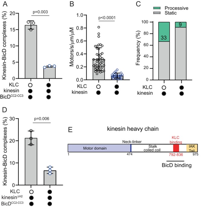
The folded auto-inhibited state of kinesin-1 is stabilized by multiple weak interactions and binds poorly to microtubules. Here we investigate the extent to which homodimeric Drosophila kinesin-1 lacking light chains is activated by the dynein activating adaptor Drosophila BicD. We show that one or two kinesins can bind to the central region of BicD (CC2), a region distinct from that which binds dynein-dynactin (CC1) and cargo-adaptor proteins (CC3). Kinesin light chain significantly reduces the amount of kinesin bound to BicD and thus regulates this interaction. Binding of BicD to kinesin enhances processive motion, suggesting that the adaptor relieves kinesin auto-inhibition. In contrast, the kinesin-binding domain of microtubule-associated protein 7 (MAP7) has minimal impact on the fraction of motors moving processively while full-length MAP7 enhances kinesin-1 recruitment to the microtubule and run length because of its microtubule-binding domain. BicD thus relieves auto-inhibition of kinesin, while MAP7 enhances motor engagement with the microtubules. When BicD and MAP7 are combined, the most robust activation of kinesin-1 occurs, highlighting the crosstalk between adaptors and microtubule-associated proteins in regulating transport.

Keywords: adaptor protein; dynein; kinesin; microtubule; processivity.


Conflict of interest statement

The authors declare no conflicts of interest.

Figures

FIGURE 1
FIGURE 1
Kinesin‐1 heavy chain binds to the central region of BicD. (A) Schematic of the series of BicD constructs used to define the kinesin binding site. Regions predicted by AlphaFold to be helical are labeled H1–H6, and the three coiled‐coil domains of BicD indicated (CC1, CC2 and CC3). The red dashed boxed (amino acids 437–535) delineates the region common to truncated constructs that bind kinesinΔH2 and move on microtubules (MTs) with an average frequency exceeding 0.06/s/μm microtubule length/μM kinesin concentration. (B) Binding frequency of moving kinesinΔH2‐BicD complexes on MTs for various BicD constructs. Moving complexes were counted from n = 26–45 MTs per construct. N = 2 experiments. (C) (Right panel) Dynein (labeled with a red Qdot) and kinesinΔH2 (labeled with a green Qdot) can simultaneously bind to BicD (yellow spots, arrows) in the presence of BicD, Egalitarian (Egl) and mRNA. (Left panel) Colocalization of dynein and kinesin was infrequently observed in the absence of BicD, Egl and mRNA. (D) Quantification of kinesin and dynein co‐localization with dynein‐dynactin and kinesin (DDK) versus dynein‐dynactin, BicD, Egl, mRNA and kinesin (DDBER‐K). Mean ± SD. N = 2 experiments, n = 29 MTs. Unpaired t test with Welch's correction, p < 0.0001.
FIGURE 2
FIGURE 2
Kinesin light chain decreases the interaction of the kinesin heavy chain with BicDCC2‐CC3. (A) Homodimeric kinesin without KLC forms more complexes with BicDCC2‐CC3 than does heterotetrameric kinesin with bound light chains (KLC) (16.4% ± 1.3% vs. 3.7% ± 0.26%, mean ± SD). Kinesin was separately expressed and purified with or without light chain. N = 3 experiments. Unpaired t‐test with Welch's correction, p = 0.003. (B) The presence of KLC decreased the number of kinesin motors that bound to the MT (0.32 ± 0.17 vs. 0.07 ± 0.03, mean ± SD). N = 2 experiments. Number of MTs analyzed was n = 41 without KLC and n = 23 with KLC. Unpaired t‐test with Welch's correction, p < 0.0001. (C) The presence of KLC reduced the fraction of motors moving processively from 33% to 9%. (D) The same experiment as in panel (A), but using kinesinΔH2 (21.3% ± 3.1% without KLC vs. 6.6% ± 1.5% with KLC, mean ± SD), N = zz experiments. Unpaired t‐test with Welch's correction, p = 0.006. A 5‐fold molar excess of purified KLC was added to a preparation of kinesinΔH2 expressed without KLC. (E) Schematic of the Drosophila kinesin heavy chain indicating that the region where the kinesin light chain (KLC) and BicD bind likely overlaps.
FIGURE 3
FIGURE 3
Two kinesins can bind to BicD. (A) The run length of kinesinΔH2 is enhanced from 1.8 ± 0.1 μm (n = 88) to 3.5 ± 0.1 μm (n = 115) in the presence of BicDCC2‐CC3, N = 3 experiments, Kolmogorov–Smirnov test, p < 0.0001. (B) Equimolar amounts of kinesinΔH2 labeled with either Alexa 488 or Alexa 647 were mixed with BicDCC2‐CC3. The speed of single‐colored images (0.52 ± 0.20, n = 88) was the same as that for dual‐colored images with 2 kinesinΔH2 bound (0.48 ± 0.17, n = 38). N = 2 experiments, unpaired t‐test with Welch's correction, p = 0.26. (C) Run length of dual‐colored images containing two kinesinΔH2 complexes is longer (3.8 ± 0.8, n = 38) than that of single‐colored images (1.8 ± 0.1, n = 88, N = 2 experiments, Kolmogorov–Smirnov test, p < 0.0001). (D) Interferometric scattering mass spectrometry (iSCAMS) data showing contrast distributions for BicDCC2‐CC3, kinesinΔH2 and the kinesinΔH2‐BicDCC2‐CC3 complex, along with a standard curve of contrast as a function of molecular mass. BicD and kinesin were each fit to a single Gaussian and the kinesin‐BicD complex histogram was fitted to the sum of two Gaussian distributions. For the single Gaussian fit (nonlinear least square fit), the coefficient of determination was R 2 = 0.966. For the two‐component Gaussian fit (nonlinear least square fit), R 2 = 0.992 and the means of the components were free and not fixed. (E) Equimolar amounts of BicDCC2‐CC3 labeled with either 525 or 655 nm Qdots were mixed with kinesinΔH2 and the number of red‐green co‐localizations quantified to show that a single kinesin does not bind to two BicD molecules. N = 2 experiments, n = 39 MTs. Unpaired t test with Welch's correction, p < 0.0001.
FIGURE 4
FIGURE 4
BicD increases the fraction of processively moving kinesin‐1 motors. (A) Kymographs illustrating the trajectories of (left) kinesin or (right) the kinesin‐BicDCC2‐CC3 complex on MTs. (B) Recruitment of kinesin (motors/s/μm/μM) (0.32 ± 0.17, n = 42 MTs), the kinesin‐BicDCC2‐CC3 complex (0.50 ± 24, n = 41 MTs) and kinesinΔH2 (0.67 ± 0.24, n = 33 MTs) to the MT. Mean ± SD, N = 3 experiments. One‐way ANOVA with Tukey's multiple comparison, p values indicated on figure. (C) Percentage of bound motors that move processively (green), diffusively (purple) or that were static (gray). The percentage of processive events were 28% for kinesin, 52% for the kinesin‐BicDCC2‐CC3 complex and 48% for kinesinΔH2. (D) Speed distributions of kinesin (0.25 ± 0.19 μm/s, n = 74), the kinesin‐BicDCC2‐CC3 complex (0.33 ± 0.19, n = 84) and kinesinΔH2 (0.43 ± 0.25 μm/s, n = 111). Mean ± SD, N = 3 experiments. One‐way ANOVA with Tukey's multiple comparison, p values indicated on figure. (E) Run length of kinesin (1.2 ± 0.1 μm, n = 34), the kinesin‐BicDCC2‐CC3 complex (2.3 ± 0.1 μm, n = 63) and kinesinΔH2 (1.9 ± 0.1 μm, n = 79). N = 3 experiments. One‐way ANOVA with Tukey's multiple comparison, p values indicated on figure.
FIGURE 5
FIGURE 5
MAP7 enhances kinesin recruitment to the MT. (A) Schematic of the kinesin‐MAP7 interaction. The kinesin‐binding domain (MAP7KBD) and the microtubule‐binding domain (MAP7MTBD) of MAP7 are indicated. (B) Kinesin was labeled with a red Qdot, and MAP7 with a green Qdot. The kinesin‐MAP7 complex can move together processively on MT tracks (yellow), but MAP7 can also dissociate during the processive run (white arrow). (C) Fields illustrating enhanced recruitment of kinesin (red) to the MT (green) in the presence of 10 nM MAP7. (D) MAP7 enhances the number of kinesin motors bound to the MT from 0.32 ± 0.17 motors/s/μm/μM to 0.86 ± 0.35 (mean ± SD, N = 2 experiments, n = 42 and 31 MTs respectively). Unpaired t‐test with Welch's correction, p < 0.0001. (E) The percentage of processive runs increases from 28% to 38% in the presence of MAP7, and the number of diffusive events increased from 5% to 13%. (F) The speed of kinesin in the presence of MAP7 (0.29 ± 0.13, n = 76) is similar to that of kinesin alone (0.25 ± 0.14, n = 61). Mean ± SD, N = 2 experiments, Unpaired t‐test with Welch's correction, p = 0.04. (G) The run length of the kinesin in the presence of MAP7 (2.5 ± 0.1 μm) is significantly longer than that of kinesin alone (1.2 ± 0.1 μm). N = 2 experiments, Kolmogorov–Smirnov test, p < 0.0001. (H) MAP7KBD did not enhance the number of kinesin motors bound to the MT (0.40 ± 0.18 motors/s/μm/μM vs. 0.47 ± 0.15, n = 12 MTs, mean ± SD, N = 2 experiments; Unpaired t‐test with Welch's correction, p = 0.32). (I) The percentage of processive runs was unchanged in the presence of MAP7KBD (27% vs. 30%). (J) The speed of kinesin in the presence of MAP7KBD (0.29 ± 0.13, n = 76) is not significantly different from that of kinesin alone (0.25 ± 0.19, n = 74). Mean ± SD, N = 2 experiments, Unpaired t‐test with Welch's correction, p = 0.30. (K) The run length of the kinesin‐MAP7KBD complex (1.2 ± 0.1 μm) was not significantly different than that of kinesin alone (1.4 ± 0.1 μm). N = 2 experiments, Kolmogorov–Smirnov test, p = 0.32.
FIGURE 6
FIGURE 6
The combination of BicD and MAP7 produces the most active kinesin. Values for the kinesin‐BicDCC2‐CC3‐MAP7 complex are shown and compared to data previously shown with other kinesin or kinesin‐complexes. (A) The combination of MAP7 and BicDCC2‐CC3 enhances the number of kinesin motors bound to microtubules (from 0.32 ± 0.17 motors/s/μm/μM to 1.1 ± 0.39, N = 3 experiments, n = 45 MTs, One‐way ANOVA with Tukey's multiple comparison. p values indicated on figure). (B) The percentage of processive runs was 62% in the presence of both MAP7 and BicDCC2‐CC3, higher than with either binding partner alone. (C) The speed of kinesin in the presence of both MAP7 and BicDCC2‐CC3 is 0.43 ± 22 μm/s, n = 67. N = 3 experiments, One‐way ANOVA with Tukey's multiple comparisons are shown with p values. (D) The run length of kinesin in the presence of both MAP7 and BicDCC2‐CC3 (3.9 ± 0.2 μm) was statistically longer than all other variations. N = 3 experiments, One‐way ANOVA with Tukey's multiple comparison.

Update of

Similar articles

Cited by

References

    1. Mohler J. and Wieschaus E. F., “Dominant Maternal‐Effect Mutations of Drosophila melanogaster Causing the Production of Double‐Abdomen Embryos,” Genetics 112 (1986): 803–822. - PMC - PubMed
    1. Olenick M. A. and Holzbaur E. L. F., “Dynein Activators and Adaptors at a Glance,” Journal of Cell Science 132 (2019): 227132. - PMC - PubMed
    1. McKenney R. J., Huynh W., Tanenbaum M. E., Bhabha G., and Vale R. D., “Activation of Cytoplasmic Dynein Motility by Dynactin‐Cargo Adapter Complexes,” Science 345 (2014): 337–341. - PMC - PubMed
    1. Schlager M. A., Hoang H. T., Urnavicius L., Bullock S. L., and Carter A. P., “In Vitro Reconstitution of a Highly Processive Recombinant Human Dynein Complex,” EMBO Journal 33 (2014): 1855–1868. - PMC - PubMed
    1. Gama J. B., Pereira C., Simões P. A., et al., “Molecular Mechanism of Dynein Recruitment to Kinetochores by the Rod‐Zw10‐Zwilch Complex and Spindly,” Journal of Cell Biology 216 (2017): 943–960. - PMC - PubMed

MeSH terms

Grants and funding

LinkOut - more resources