. 2025 Jun;51(3):e70019.
doi: 10.1111/nan.70019.

Alpha-Synuclein as a Potential Biomarker for Inclusion Body Myositis in Blood and Muscle

Affiliations

Alpha-Synuclein as a Potential Biomarker for Inclusion Body Myositis in Blood and Muscle

Tobias Mayer et al. Neuropathol Appl Neurobiol. 2025 Jun.

Abstract

Aims: Diagnosis of inclusion body myositis (IBM) is difficult and currently based on a combination of clinical and (immuno)histological findings. Biomarkers facilitating the diagnostic process are needed. Alpha-synuclein (αSN) aggregates are a known histological feature of IBM, but there is a lack of information on their diagnostic relevance. Furthermore, serum αSN concentrations in IBM have not been investigated.

Methods: Immunohistochemical staining for αSN was performed on 63 biopsies (19 IBM, 21 other inflammatory myopathies, 20 other myopathies and 3 healthy controls), and αSN reactive fibres were quantified. The serum concentration of αSN was determined by ELISA in 156 serum samples (11 IBM, 25 other inflammatory myopathies, 53 hereditary myopathies, 30 mitochondriopathies and 37 healthy controls).

Results: The proportion of fibres with αSN immunoreactivity was significantly higher in IBM compared to all groups (p < 0.001) and discriminated IBM against all other neuromuscular disorders with a sensitivity of 79% and a specificity of 85%, which further improved when only non-regenerating fibres were examined. In serum, αSN concentrations in IBM were generally not different from healthy controls. However, serum concentrations were inversely correlated with disease duration (r = -0.62, p = 0.04) and positively correlated with the IBM functional rating scale (r = 0.74, p = 0.01). Consequently, stratification according to these clinical parameters showed significantly lower serum αSN concentrations in late-stage, more severely affected patients.

Conclusions: αSN reactivity may serve as an additional immunohistochemical marker for IBM diagnosis. Furthermore, this study indicates that αSN serum concentrations decrease with disease duration and clinical deterioration. Therefore, serum αSN may be provisionally considered a monitoring biomarker in IBM, pending further studies.

Keywords: IBM; biomarker; inclusion body; myositis; α‐synuclein.


Conflict of interest statement

The authors declare no conflicts of interest.

Figures
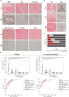
FIGURE 1
FIGURE 1
Distribution and quantification of αSN immunoreactivity. (a) HE and αSN staining. IBM samples exhibited strong immunoreactivity for αSN, while only single fibres were positive in other forms of IIM and myopathies with rimmed vacuoles. Note the subsarcolemmal enhancement of some fibres in IBM (black arrowheads). Necrotic fibres did not show reactivity (white arrowhead). (b) Allocation of αSN reactivity in different fibre types. Most fibres in IBM showed small dotty inclusions (DOT), while less pronounced, homogeneous reactivity (REG) was found in most regenerating fibres. No reactivity was observed within rimmed vacuoles (RV). (c) Nature of immunoreactive fibres in different groups. In samples other than IBM, regenerating fibres accounted for a larger proportion of immunoreactive fibres. (d) Quantification of αSN immunoreactivity and ROC analysis to differentiate IBM from the other subgroups studied. All fibres: Quantification revealed a significantly higher proportion of reactive fibres (%) in IBM samples, while ROC analysis shows a good differentiation of IBM from other IIMs and other myopathies. Non–regenerating fibres: Quantification of αSN immunoreactivity only in non–regenerating fibres improved discrimination of IBM from other IIMs. The discrimination of IBM from other myopathies in general and myopathies with rimmed vacuoles did not differ after excluding regenerating fibres. The ROC analyses of IBM versus VAC (non‐rim.), non‐VAC and CTR yielded a sensitivity and specificity of 100%, respectively. These results are not displayed here to enhance clarity. p < 0.05*, p < 0.01**, p < 0.001*** and p < 0.0001****. Abbreviations: all, all specimens other than IBM; CTR, control specimens; DOT, dotty αSN immunoreactivity; HE, haematoxylin eosin staining; IBM, inclusion body myositis; IIM, idiopathic inflammatory myopathies; NMD, neuromuscular diseases (including IIM and other myopathies); REG, regenerating muscle fibre; RV, rimmed vacuole; VAC (rim.), vacuolar myopathies with rimmed vacuoles; VAC (non‐rim.), vacuolar myopathies with non–rimmed vacuoles; non‐VAC, other myopathies without vacuoles; αSN, α‐synuclein staining; scale bars: (a) 50 μm and (b) 20 μm.
FIGURE 2
FIGURE 2
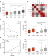
Analysis of αSN concentration in serum samples. (a) IBM samples tended to have lower serum αSN concentration than other groups (left side, notice median), although not significant. (b) Correlation of serum αSN concentration with different epidemiological and clinical parameters. Serum αSN concentration correlated significantly with patients' disease duration and IBMFRS. IBMFRS correlated significantly with CK activity. (c) Correlation analysis of αSN with patients' disease duration (left) and stratification of IBM patients in an early (≤ 8 years disease duration) and a late‐stage (> 8 years disease duration) IBM (right). (d) Correlation analysis of αSN with patients' IBMFRS (left) and stratification in mild (IBMFRS > 20) and advanced (IBMFRS ≤ 20) IBM (right side). Abbreviations: A‐IBM, advanced IBM; CK, creatine kinase; CTR, control samples; E‐IBM, early‐stage IBM; HER, hereditary myopathies; IBMFRS, inclusion body myositis functional rating scale; IBM, inclusion body myositis; IIM, idiopathic inflammatory myopathies; L‐IBM, late‐stage IBM; M‐IBM, mild IBM; MIT, mitochondrial myopathies; modified MRC sum score, modified Medical Research Council sum score; αSN, α‐synuclein.

References

    1. Lundberg I. E., Fujimoto M., Vencovsky J., et al., “Idiopathic Inflammatory Myopathies,” Nature Reviews. Disease Primers 7, no. 1 (2021): 86. - PubMed
    1. Hilton‐Jones D. and Brady S., “Diagnostic Criteria for Inclusion Body Myositis,” Journal of Internal Medicine 280, no. 1 (2016): 52–62. - PubMed
    1. Greenberg S. A., “Inclusion Body Myositis: Clinical Features and Pathogenesis,” Nature Reviews Rheumatology 15, no. 5 (2019): 257–272, 10.1038/s41584-019-0186-x. - DOI - PubMed
    1. Cox F. M., Verschuuren J. J., Verbist B. M., Niks E. H., Wintzen A. R., and Badrising U. A., “Detecting Dysphagia in Inclusion Body Myositis,” Journal of Neurology 256, no. 12 (2009): 2009–2013. - PMC - PubMed
    1. Lucchini M., Maggi L., Pegoraro E., et al., “Anti‐cN1A Antibodies Are Associated With More Severe Dysphagia in Sporadic Inclusion Body Myositis,” Cells 10, no. 5 (2021): 1146, 10.3390/cells10051146. - DOI - PMC - PubMed

Publication types

LinkOut - more resources