. 2025 Aug;292(16):4272-4288.
doi: 10.1111/febs.70130. Epub 2025 May 19.

Genomic context analysis enables the discovery of an unusual NAD-dependent racemase in phosphonate catabolism

Affiliations

Genomic context analysis enables the discovery of an unusual NAD-dependent racemase in phosphonate catabolism

Francesca Ruffolo et al. FEBS J. 2025 Aug.

Abstract

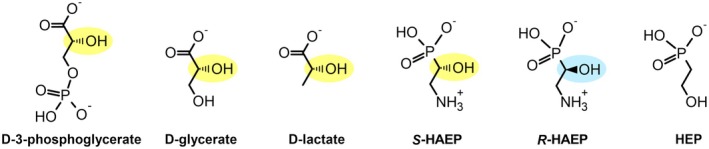
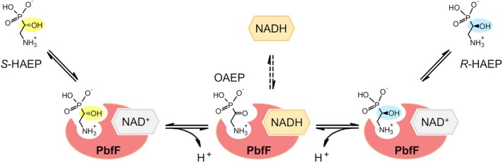
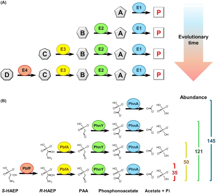
Phosphonates are organic molecules containing a direct carbon-phosphorus (C-P) bond. They are chemically sturdy compounds that can, however, be degraded by environmental microorganisms. In the frame of bacterial phosphonate catabolism, we recently reported the discovery of (R)-1-hydroxy-2-aminoethylphosphonate ammonia-lyase (PbfA), a lyase acting on the natural compound (R)-2-amino-1-hydroxyethylphosphonate (R-HAEP). PbfA converts R-HAEP into phosphonoacetaldehyde (PAA), which can be subsequently processed and cleaved by further enzymes. However, PbfA is not active toward S-HAEP (the enantiomer of R-HAEP), whose metabolic fate remained unknown. We now describe the identification of a racemase, discovered through genomic context analysis, which converts S-HAEP into R-HAEP, thereby enabling degradation of S-HAEP. We propose for this enzyme the official name 2-amino-1-hydroxyethylphosphonate racemase (shorthand PbfF). To our knowledge, PbfF is the first NAD-dependent racemase ever described and is structurally unrelated to other known NAD-dependent isomerases. The enzyme uses NAD+ as a cofactor, is inhibited by NADH, and shows catalytic parameters comparable to those of other racemases acting on similar substrates. The presence of a pathway for the breakdown of S-HAEP in numerous bacteria suggests that this compound may be more common in the environment than currently appreciated. Notably, the route for S-HAEP degradation appears to have developed through a mechanism of retrograde metabolic evolution.

Keywords: 2‐amino‐1‐hydroxyethylphosphonate; Alphaproteobacteria; NAD‐dependent isomerase; phosphonate degradation; racemase.


Conflict of interest statement

The authors declare no conflict of interest.

Figures

Fig. 1
Fig. 1
Hydrolytic pathways for the microbial catabolism of 2‐aminoethylphosphonate (AEP) and of some related compounds. (A) For AEP, the first step is the conversion to phosphonoacetaldehyde (PAA), typically operated by the transaminase PhnW (or more rarely by an amine oxidase; not shown [22]). PAA can then be hydrolyzed into acetaldehyde and phosphate by PhnX (upper route; PhnWX pathway), or, alternatively, it can be first converted to phosphonoacetate and then to acetate and phosphate by two consecutive reactions, catalyzed by the enzymes PhnY and PhnA (lower route; PhnWYA pathway). (B) The natural compound R‐HAEP can also be converted to PAA through the action of the lyase PbfA [21]. (C) The N‐monomethylated form of AEP (M1AEP) generates PAA through an oxidative deamination, which can be catalyzed by a FAD‐dependent dehydrogenase (PbfC) or by a FAD‐dependent oxidase (PbfD) [22].
Fig. 2
Fig. 2
Genes coding for a predicted NAD‐dependent dehydrogenase (PbfF) in clusters dedicated to AEP degradation. The pbfF genes are shown in red; the accession IDs of the encoded proteins are provided in Table 1. Other highlighted genes include phnW (light purple), phnA (azure), phnY (green), phnX (olive), pbfA (yellow), pbfC (ice blue) and pbfD (orange). Putative transporter genes are shown in light gray, whereas predicted transcription regulators are in dark gray.
Fig. 3
Fig. 3
Structural comparison between the substrates of d‐hydroxyacid dehydrogenases and potential substrates of PbfF. The d‐hydroxyacids are shown on the left and some naturally occurring hydroxy‐containing phosphonates are shown on the right. Chiral centers, when present, are highlighted in yellow or light blue.
Fig. 4
Fig. 4
A phosphate release assay shows the involvement of PbfF in the degradation of S‐HAEP. Different phosphonates (S‐HAEP, R‐HAEP or HEP, 3 mm) were tested as described in the Methods. PbfF, in combination with PbfA and PhnX, afforded phosphate release from S‐HAEP. The presence of both PbfF and PbfA was essential: as much as PbfA alone is unable to process S‐HAEP [21], PbfF, in the absence of PbfA, could not accomplish the degradation of S‐HAEP (nor of R‐HAEP). The experiment was replicated three times with essentially identical results.
Fig. 5
Fig. 5
Accumulation of NADH upon incubation of PbfF with HAEP isomers. Each kinetic trace is representative of at least three repetitions. (A) Reaction of PbfF with S‐HAEP. The enzyme (0.9 μm) to a solution containing 3 mmS‐HAEP and 0.3 mm NAD+ in 50 mm Bis‐Tris propane at three different pH values, 23 °C. The amount of NADH formed was calculated based on the increase of absorbance at 340 nm. At pH 8 NADH formation was almost undetectable. Even at pH 10 (where NAD+ reduction is intrinsically favored) the NADH accumulated in 15 min was <10% of the total NAD. (B) Reaction of PbfF with R‐HAEP. Apart from the presence of 3 mm R‐HAEP (in place of S‐HAEP) other conditions were as in panel A. (C) Effect of the addition of PbfA on the accumulation of NADH upon reaction of PbfF with S‐HAEP. The reaction was conducted as in panel A (pH 10.0) except that after about 28 min the reaction mixture was supplemented with 2 μm PbfA.
Fig. 6
Fig. 6
Circular dichroism (CD) assessment of the racemase activity of PbfF. Each reported experiment is representative of three repetitions. (A) CD spectra of S‐HAEP and R‐HAEP. Solutions of the two enantiomers (5 mm in each case, pH 8.0) were placed in a 1 mL quartz cuvette with 0.5 cm path length and spectra were collected using a J‐1500 CD spectrophotometer (Jasco Inc., Easton, MD, USA). Bandwidth 1 nm, scanning speed 50 nm·min−1. (B) CD signals of the HAEP enantiomers disappear upon PbfF treatment. Solutions initially containing S‐HAEP (5 mm, pH 8.0) and NAD+ (50 μm) were incubated for 75 min in the absence (red line) or in the presence (blue line) of 1 μm PbfF. Solutions with R‐HAEP and NAD+ were also incubated for 75 min in the absence (green line) or in the presence (yellow line) of 1 μm PbfF. All solutions were ultrafiltered before collecting the spectra. (C) UV absorption spectra of the same samples as in panel B show no disappearance of chromophoric species due to PbfF. The band at ~260 nm and the shoulder at ~215 nm are attributable to NAD+.
Fig. 7
Fig. 7
PbfF activity as a function of the concentrations of cofactor and of substrate (50 mm TEA‐HCl, pH 8.0, 25 °C). (A) Dependence of PbfF activity on exogenous NAD+. In addition to PbfF (35 nm) the reaction mixture contained 1 mmS‐HAEP, 5 mm MgCl2, 7.7 μm PbfA, 3.6 μm PhnX, 6.66 U·mL−1 ADH, and 0.3 mm NADPH. For each NAD+ concentration tested, two technical replicates were collected (gray circles). The orange circles show the activity in the presence of 1 mm NAD+ and 60 mm NADH. (B) Dependence of activity on the concentration of S‐HAEP. Conditions were as in panel A, except that NAD+ was kept at 1 mm while the concentration of S‐HAEP varied from 0.05 to 1.5 mm. Data points represent the average (±SD) of three technical replicates.
Fig. 8
Fig. 8
Predicted structure of PbfF from M. plurifarium and comparison with the experimental crystal structure of a pfam02826 dehydrogenase. (A) Three‐dimensional structure of the PbfF monomer predicted by AlphaFold 3 [39]. The bound NAD+ cofactor is shown in ball‐and‐stick. (B) Structure of d‐lactate dehydrogenase from P. aeruginosa (PDB: 5Z20) [30]. (C) Structural alignment of the two structures in panels A and B, generated by PyMOL [41]. (D) Schematic representation of the interactions formed by NAD+ bound to the predicted PbfF structure. The interactions were visualized through the PoseEdit [42] web tool (https://proteins.plus). (E) Interactions formed by NADH in the active site of P. aeruginosa d‐lactate dehydrogenase. Several of the interactions match closely the predicted interactions at the PbfF active site (panel D). Some of the differences (e.g., the lack of an ionic interaction with the phosphate groups in 5Z20) might be related to the different functions of the two proteins (dehydrogenase vs. isomerase).
Fig. 9
Fig. 9
Tentative scheme for the reaction catalyzed by PbfF. The conversion of S‐HAEP into R‐HAEP proceeds without net production of NADH. However, NADH is formed transiently as an enzyme‐bound intermediate, which is expectedly more stable at high pH (NAD+ reduction is accompanied by the release of one proton). Over time, some NADH may also leak out of the active site (dashed vertical arrows).
Fig. 10
Fig. 10
‘Retrograde’ emergence of the S‐HAEP degradation route. (A) The retrograde model of metabolic pathway evolution [60] posits that evolution of a pathway begins with a first enzyme (E1) able to transform an environmental compound A to yield a useful product P. The progressive depletion of A leads to a selective pressure for the development of a second enzyme (E2), which can obtain A from an available precursor B. When B becomes depleted, yet another enzyme (E3) can emerge, and so forth. (B) Development of the S‐HAEP degradation pathway. Numbers on the right refer to a census of phnA genes in 1098 complete genomes of Hyphomicrobiales and Rhodobacterales, annotated as “finished” in the IMG/M website.

Similar articles

  • Discovery of a New, Recurrent Enzyme in Bacterial Phosphonate Degradation: (R)-1-Hydroxy-2-aminoethylphosphonate Ammonia-lyase.
    Zangelmi E, Stanković T, Malatesta M, Acquotti D, Pallitsch K, Peracchi A. Zangelmi E, et al. Biochemistry. 2021 Apr 20;60(15):1214-1225. doi: 10.1021/acs.biochem.1c00092. Epub 2021 Apr 8. Biochemistry. 2021. PMID: 33830741 Free PMC article.
  • Prescription of Controlled Substances: Benefits and Risks.
    Preuss CV, Kalava A, King KC. Preuss CV, et al. 2025 Jul 6. In: StatPearls [Internet]. Treasure Island (FL): StatPearls Publishing; 2025 Jan–. 2025 Jul 6. In: StatPearls [Internet]. Treasure Island (FL): StatPearls Publishing; 2025 Jan–. PMID: 30726003 Free Books & Documents.
  • Sexual Harassment and Prevention Training.
    Cedeno R, Bohlen J. Cedeno R, et al. 2024 Mar 29. In: StatPearls [Internet]. Treasure Island (FL): StatPearls Publishing; 2025 Jan–. 2024 Mar 29. In: StatPearls [Internet]. Treasure Island (FL): StatPearls Publishing; 2025 Jan–. PMID: 36508513 Free Books & Documents.
  • Citrullinemia Type I.
    Quinonez SC, Lee KN. Quinonez SC, et al. 2004 Jul 7 [updated 2022 Aug 18]. In: Adam MP, Feldman J, Mirzaa GM, Pagon RA, Wallace SE, Amemiya A, editors. GeneReviews® [Internet]. Seattle (WA): University of Washington, Seattle; 1993–2025. 2004 Jul 7 [updated 2022 Aug 18]. In: Adam MP, Feldman J, Mirzaa GM, Pagon RA, Wallace SE, Amemiya A, editors. GeneReviews® [Internet]. Seattle (WA): University of Washington, Seattle; 1993–2025. PMID: 20301631 Free Books & Documents. Review.
  • Signs and symptoms to determine if a patient presenting in primary care or hospital outpatient settings has COVID-19.
    Struyf T, Deeks JJ, Dinnes J, Takwoingi Y, Davenport C, Leeflang MM, Spijker R, Hooft L, Emperador D, Domen J, Tans A, Janssens S, Wickramasinghe D, Lannoy V, Horn SRA, Van den Bruel A; Cochrane COVID-19 Diagnostic Test Accuracy Group. Struyf T, et al. Cochrane Database Syst Rev. 2022 May 20;5(5):CD013665. doi: 10.1002/14651858.CD013665.pub3. Cochrane Database Syst Rev. 2022. PMID: 35593186 Free PMC article.

References

    1. Horsman GP & Zechel DL (2017) Phosphonate biochemistry. Chem Rev 117, 5704–5783. - PubMed
    1. Hilderbrand RL & Henderson T (1983) Phosphonic acids in nature. In The Role of Phosphonates in Living Systems (Hilderbrand RL, ed.), pp. 5–28. CRC Press, Boca Raton, FL, USA.
    1. Kafarski P (2019) Phosphonates: their natural occurrence and physiological role. In Contemporary Topics about Phosphorus in Biology and Materials (Churchill DG, Sikirić MD, Čolović B & Milhofer HF, eds), pp. 1–19. Intech Open, London, UK.
    1. Li S & Horsman GP (2022) An inventory of early branch points in microbial phosphonate biosynthesis. Microb Genom 8, 000781. - PMC - PubMed
    1. Rott E, Steinmetz H & Metzger JW (2018) Organophosphonates: a review on environmental relevance, biodegradability and removal in wastewater treatment plants. Sci Total Environ 615, 1176–1191. - PubMed

MeSH terms

LinkOut - more resources