

Experimental methods for studying amyloid cross-interactions
- PMID: 40384558
- PMCID: PMC12086524
- DOI: 10.1002/pro.70151
Experimental methods for studying amyloid cross-interactions
Abstract
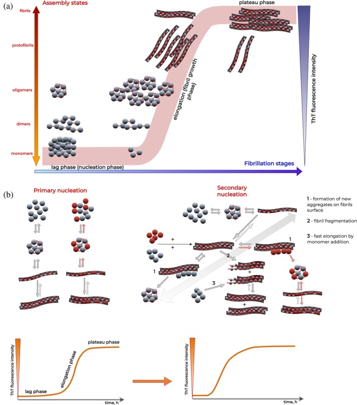
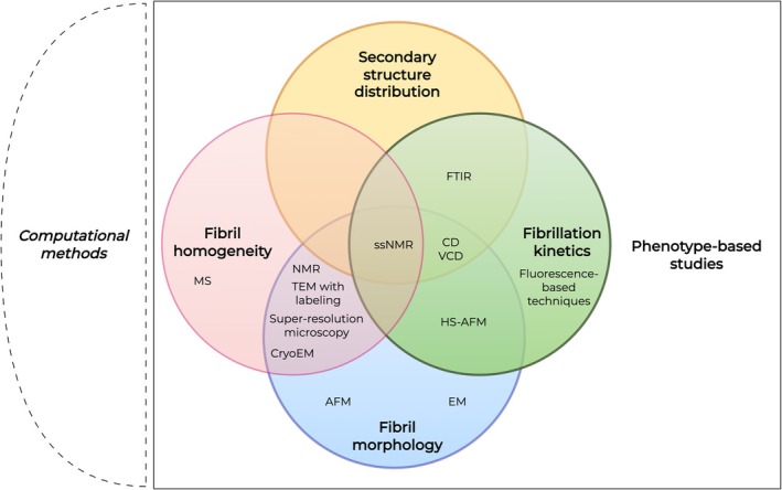
Interactions between amyloid proteins represent the cornerstone of various pathogenic pathways, including prion conversion and co-development of distinct kinds of systemic amyloidosis. Various experimental methodologies provide insights into the effects of such cross-interactions on amyloid self-assembly, which range from acceleration to complete inhibition. Here, we present a comprehensive review of experimental methods most commonly used to study amyloid cross-interactions both in vitro and in vivo, such as fluorescence-based techniques, high-resolution imaging, and spectroscopic methods. Although each method provides distinct information on amyloid interactions, we highlight that no method can fully capture the complexity of this process. In order to achieve an exhaustive portrayal, it is necessary to employ a hybrid strategy combining different experimental techniques. A core set of fluorescence methods (e.g., thioflavin T) and high-resolution imaging techniques (e.g., atomic force microscopy or Cryo-EM) are required to verify the lack of self-assembly or alterations in fibril morphology. At the same time, immuno-electron microscopy, mass spectrometry, or solid-state NMR can confirm the presence of heterotypic fibrils.
Keywords: cross‐interactions; cross‐seeding; fibril polymorphism; high‐resolution microscopy; thioflavin T.
© 2025 The Author(s). Protein Science published by Wiley Periodicals LLC on behalf of The Protein Society.
Conflict of interest statement
The authors report there are no competing interests to declare.
Figures
Similar articles
-
Structural Identification of Individual Helical Amyloid Filaments by Integration of Cryo-Electron Microscopy-Derived Maps in Comparative Morphometric Atomic Force Microscopy Image Analysis.J Mol Biol. 2022 Apr 15;434(7):167466. doi: 10.1016/j.jmb.2022.167466. Epub 2022 Jan 22. J Mol Biol. 2022. PMID: 35077765 Free PMC article.
-
Methods for Structural Analysis of Amyloid Fibrils in Misfolding Diseases.Methods Mol Biol. 2019;1873:109-122. doi: 10.1007/978-1-4939-8820-4_7. Methods Mol Biol. 2019. PMID: 30341606
-
Amyloid-type Protein Aggregation and Prion-like Properties of Amyloids.Chem Rev. 2021 Jul 14;121(13):8285-8307. doi: 10.1021/acs.chemrev.1c00196. Epub 2021 Jun 17. Chem Rev. 2021. PMID: 34137605 Review.
-
Inhibition of lysozyme amyloidogenesis by phospholipids. Focus on long-chain dimyristoylphosphocholine.Biochim Biophys Acta Gen Subj. 2017 Nov;1861(11 Pt A):2934-2943. doi: 10.1016/j.bbagen.2017.08.023. Epub 2017 Sep 1. Biochim Biophys Acta Gen Subj. 2017. PMID: 28865760
-
Single-molecule observation of self-propagating amyloid fibrils.Microscopy (Oxf). 2022 Jun 6;71(3):133-141. doi: 10.1093/jmicro/dfac011. Microscopy (Oxf). 2022. PMID: 35253856 Review.
References
-
- Adamcik J, Mezzenga R. Study of amyloid fibrils via atomic force microscopy. Curr Opin Colloid Interface Sci. 2012;17:369–376. 10.1016/j.cocis.2012.08.001 - DOI
-
- Adil LR, Ramakrishnan V. Assessment of amyloid morphology using electron microscopy. In: Ramakrishnan V, editor. Biophysical Characterization of Functional Peptides. New York, NY: Springer US; 2023. p. 129–133. 10.1007/978-1-0716-3405-9_19 - DOI
-
- Akbarian M, Kianpour M, Yousefi R, Moosavi‐Movahedi AA. Characterization of insulin cross‐seeding: the underlying mechanism reveals seeding and denaturant‐induced insulin fibrillation proceeds through structurally similar intermediates. RSC Adv. 2020;10:29885–29899. 10.1039/D0RA05414C - DOI - PMC - PubMed
Publication types
MeSH terms
Substances
Grants and funding
LinkOut - more resources
Full Text Sources
