

Enhancing pediatric abdominal pain diagnosis: the role of ultrasound layered scanning technique
- PMID: 40384668
- PMCID: PMC12084742
- DOI: 10.21037/qims-24-1855
Enhancing pediatric abdominal pain diagnosis: the role of ultrasound layered scanning technique
Abstract
Background: Pediatric abdominal pain is a common yet diagnostically challenging symptom, particularly in young children who struggle to articulate their discomfort. With obesity increasingly affecting ultrasound accuracy, this study aimed to find the cause of pediatric abdominal pain by seeking new approaches and methods in ultrasound examination, especially in the application among obese or overweight pediatric patients.
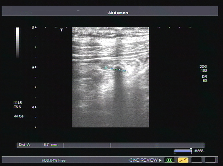
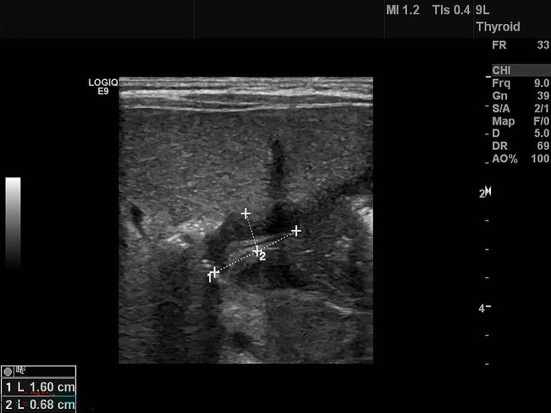
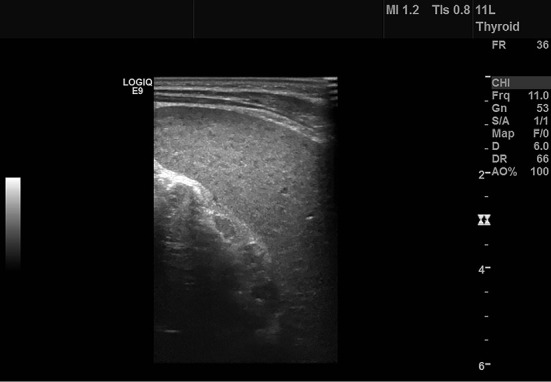
Methods: A retrospective analysis was conducted on pediatric patients hospitalized between July 2016 and November 2017 for abdominal pain. Patients were categorized into normal weight, overweight, and obese groups. Conventional and layer-by-layer scanning methods were used by attending physicians to examine abdominal organs, including the liver, gallbladder, spleen, pancreas, kidneys, and bladder. An abdominal probe was employed for rapid screening, followed by a high-frequency probe for detailed three-layer scanning. Ultrasound images were analyzed alongside the children's symptoms and physical signs to provide diagnostic insights.
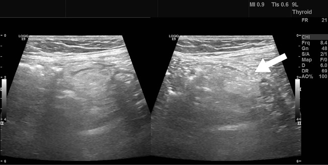
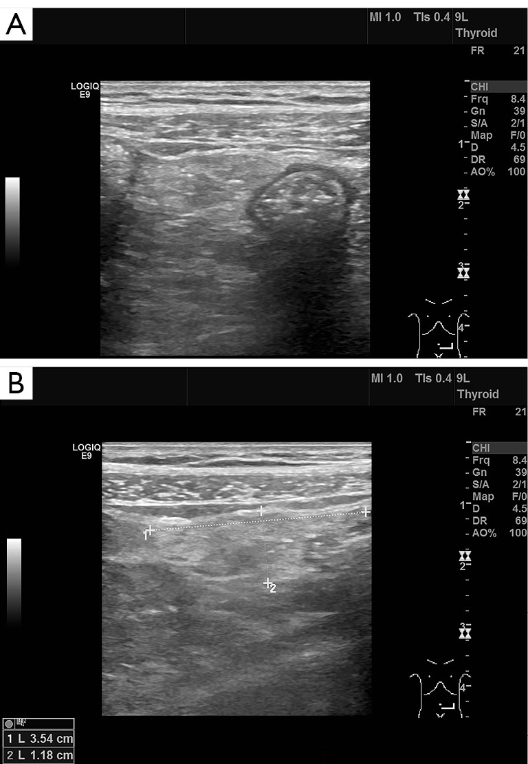
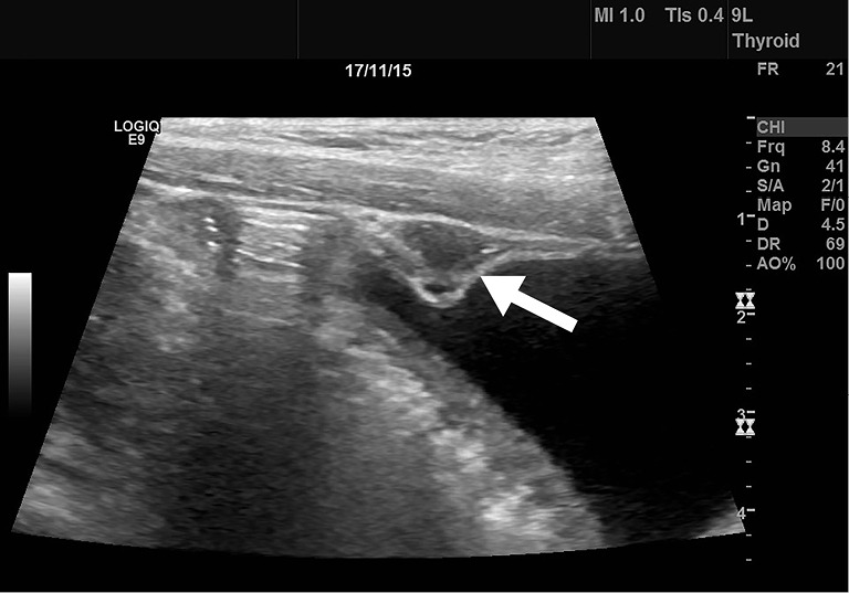
Results: When comparing the conventional and stratified screening groups, several key differences were noted. The stratified group had higher detection rates for mesenteric lymphadenopathy (100% vs. 86%) and peritonitis (94% vs. 27%). Improved detection in the stratified group was due to the identification of peritoneal thickening, leading to higher detection rates for mesenteric fat inflammation (100% vs. 46%), appendicitis (94% vs. 63%), and urachal inflammation (100% vs. 0%). Detection rates for substantial lesions, such as gallstones and ovarian torsion, were similar in both groups (100%). The stratified group also showed significantly better detection of gastrointestinal conditions like gastroenteritis (97% vs. 32%), inguinal hernia (100% vs. 0%), and intestinal ascariasis (100% vs. 47%). Differences in detection rates were observed when abdominal fat layer thickness was between 0.8 and 1.7 cm, with more significant differences when thickness exceeded 1.7 cm.
Conclusions: Real-time ultrasound with stratified screening effectively detects abdominal and pelvic masses, solid organ lesions, and bowel wall thickening, improving disease detection in children, including individuals with increased body mass index. This method is valuable and recommended for wider use.
Keywords: Abdominal pain; high resolution; layered scanning; pediatrics; real-time ultrasound.
Copyright © 2025 AME Publishing Company. All rights reserved.
Conflict of interest statement
Conflicts of Interest: All authors have completed the ICMJE uniform disclosure form (available at https://qims.amegroups.com/article/view/10.21037/qims-24-1855/coif). The authors have no conflicts of interest to declare.
Figures
Similar articles
-
Vesicoureteral Reflux.2024 Apr 30. In: StatPearls [Internet]. Treasure Island (FL): StatPearls Publishing; 2025 Jan–. 2024 Apr 30. In: StatPearls [Internet]. Treasure Island (FL): StatPearls Publishing; 2025 Jan–. PMID: 33085409 Free Books & Documents.
-
[Ultrasonography in acute pelvic pain].Acta Med Croatica. 2002;56(4-5):171-80. Acta Med Croatica. 2002. PMID: 12768897 Review. Croatian.
-
Visceral adiposity and inflammatory bowel disease.Int J Colorectal Dis. 2021 Nov;36(11):2305-2319. doi: 10.1007/s00384-021-03968-w. Epub 2021 Jun 9. Int J Colorectal Dis. 2021. PMID: 34104989 Review.
-
Faecal retention: a common cause in functional bowel disorders, appendicitis and haemorrhoids--with medical and surgical therapy.Dan Med J. 2015 Mar;62(3):B5031. Dan Med J. 2015. PMID: 25748875
-
Clinicopathological features and surgical procedures of adnexal masses with abdominal pain in pediatric and adolescent patients.Orphanet J Rare Dis. 2024 Mar 21;19(1):132. doi: 10.1186/s13023-024-03101-4. Orphanet J Rare Dis. 2024. PMID: 38515195 Free PMC article.
References
-
- Zhao XW. Discussion on pediatric emergency medicine model. Chin J Appl Clin Pediatr 2000;15:2.
-
- Brau F, Papin M, Batard E, Abet E, Frampas E, Le Thuaut A, Montassier E, Le Bastard Q, Le Conte P. Impact of emergency physician performed ultrasound in the evaluation of adult patients with acute abdominal pain: a prospective randomized bicentric trial. Scand J Trauma Resusc Emerg Med 2024;32:15. 10.1186/s13049-024-01182-5 - DOI - PMC - PubMed
LinkOut - more resources
Full Text Sources