Case Reports
. 2025 May 1;15(5):4796-4815.
doi: 10.21037/qims-24-2273. Epub 2025 Apr 17.

Transverse venous sinus stenting for fulminant idiopathic intracranial hypertension during pregnancy: a report of two cases and literature review

Affiliations
Case Reports

Transverse venous sinus stenting for fulminant idiopathic intracranial hypertension during pregnancy: a report of two cases and literature review

Tamir Regev et al. Quant Imaging Med Surg. .

Abstract

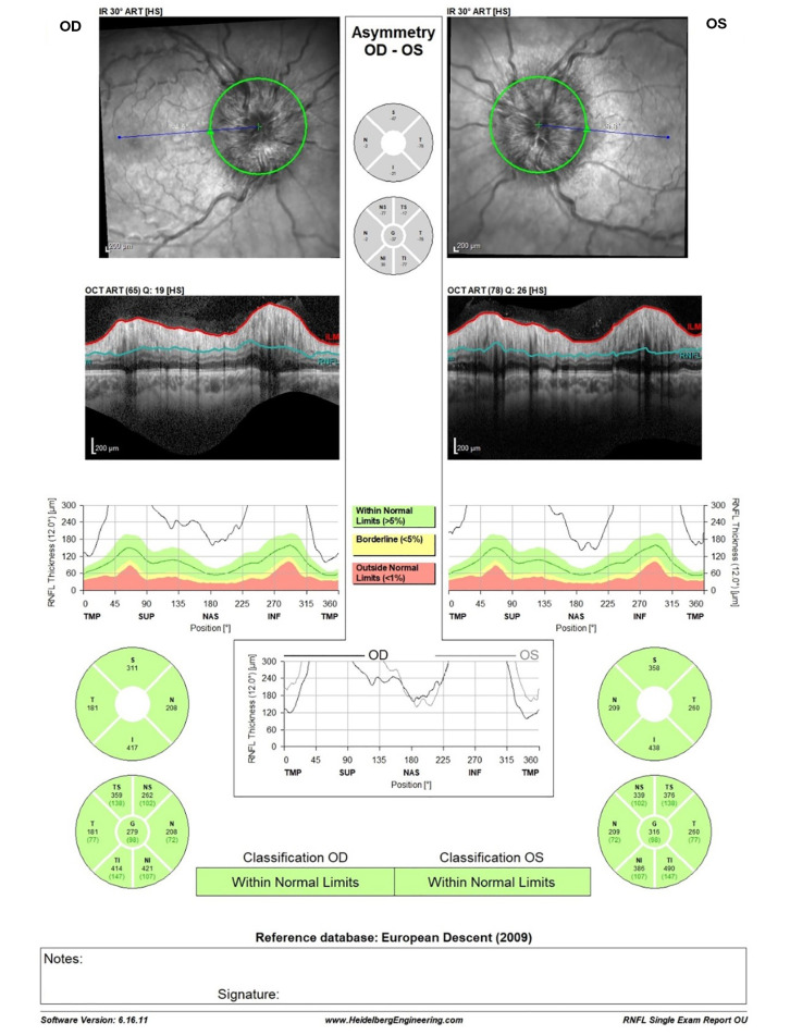
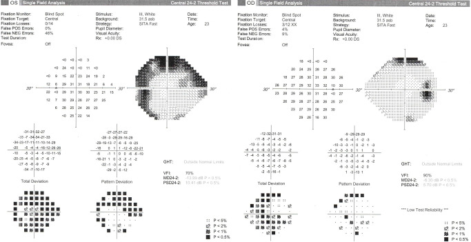
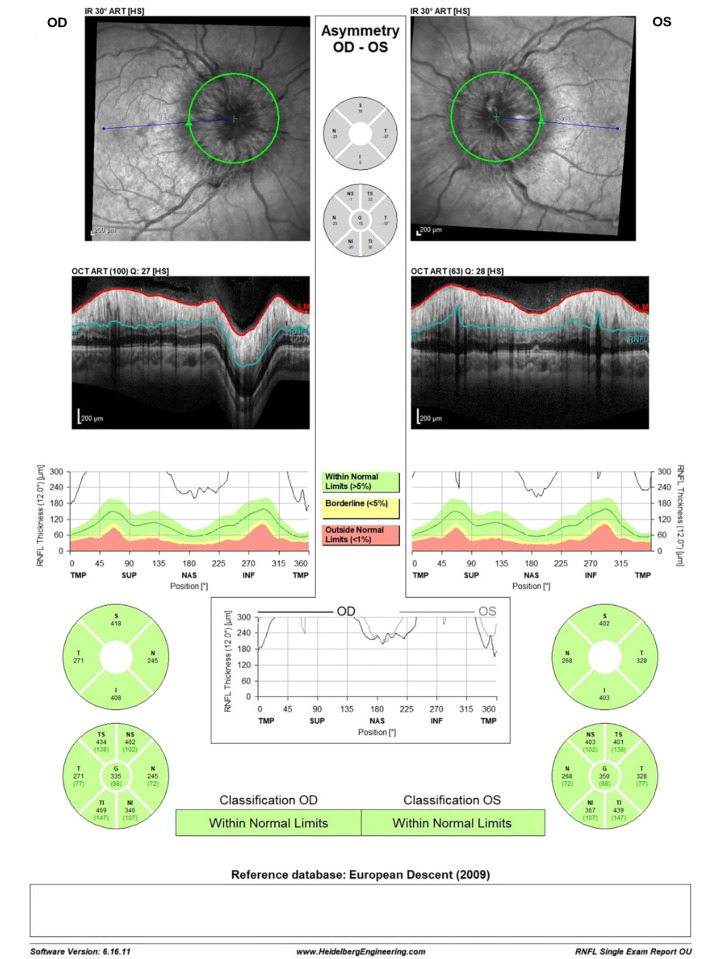
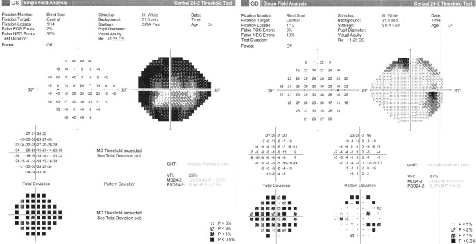
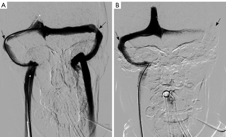
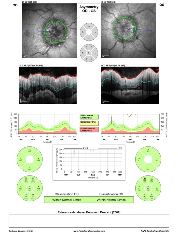
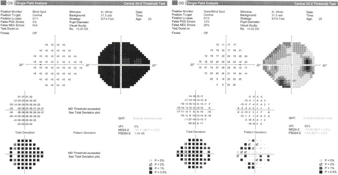
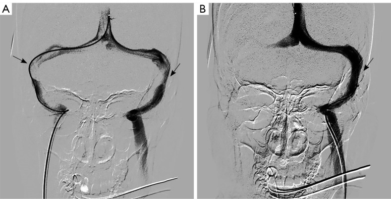
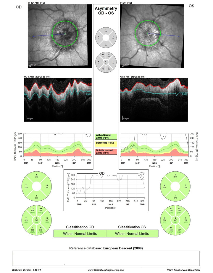
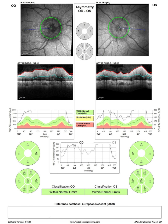
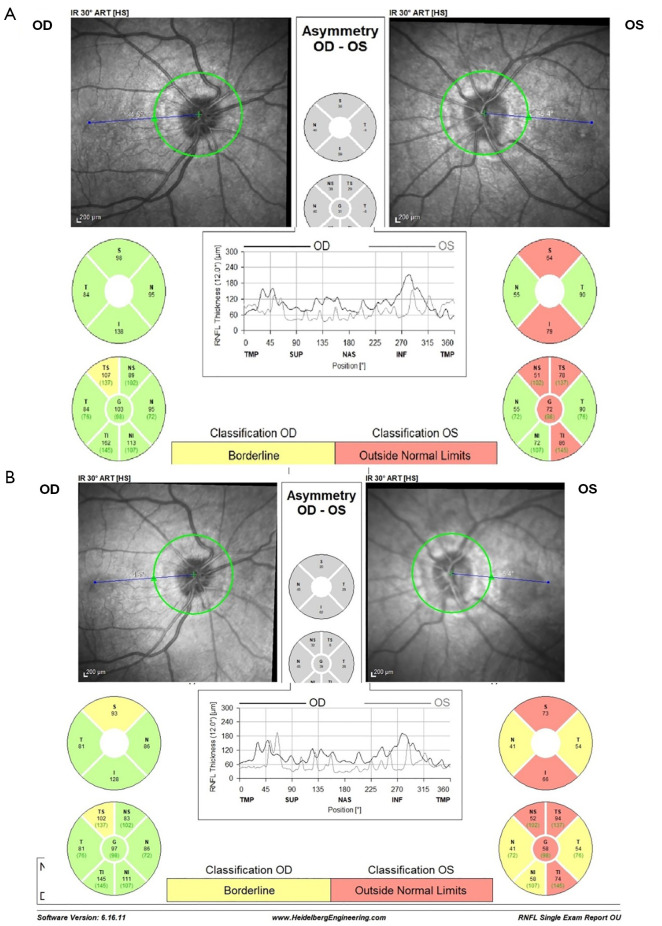
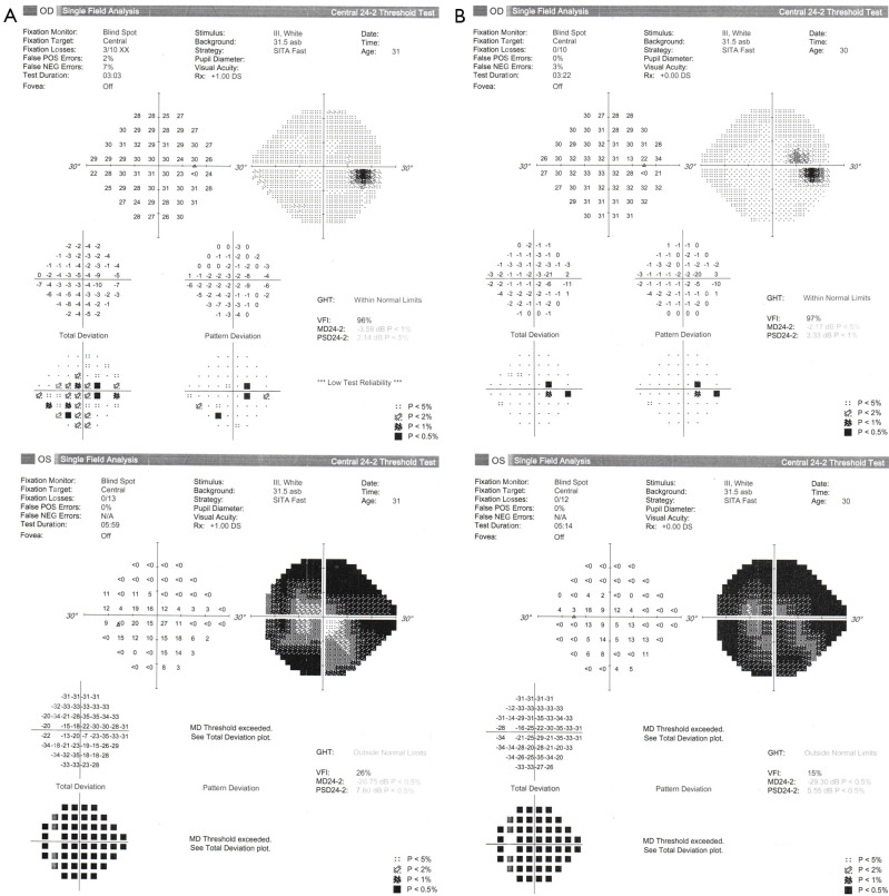
Background: Fulminant idiopathic intracranial hypertension (FIIH) is a rapidly progressive condition that carries a high risk of permanent vision loss, and is thus considered a medical emergency requiring immediate intervention. Cerebral venous sinus stenting (VSS) has gained recognition in recent years as an effective treatment for idiopathic intracranial hypertension (IIH) in patients resistant or intolerant to medical therapy. The advantage of VSS over other surgical options is its less invasive nature (endovascular procedure) and its potential to yield improvement of additional disease symptoms, such as headaches and pulsatile tinnitus. While growing evidence supports the safety and efficacy of VSS in elective cases, data on its outcomes when performed emergently in FIIH is limited and even scarcer regarding its use during pregnancy.

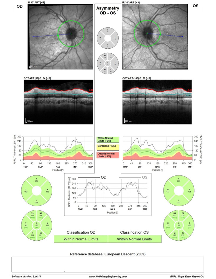
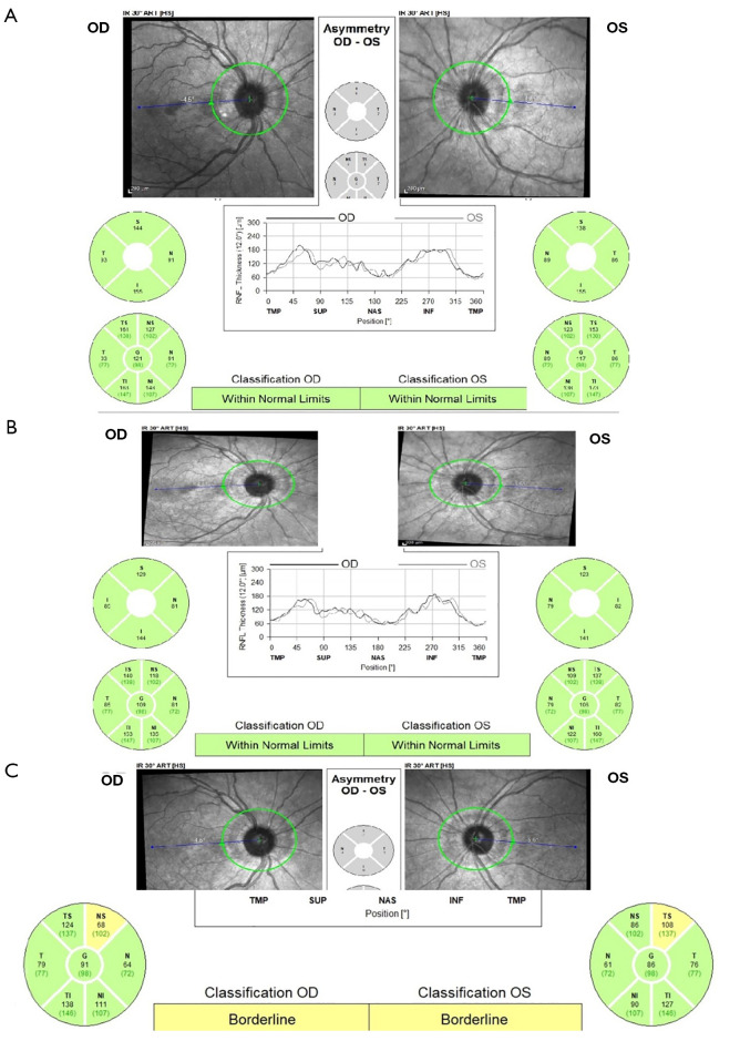
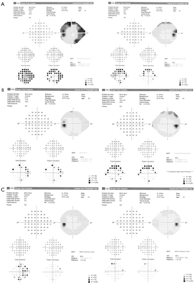
Case description: This is a retrospective description of two pregnant women in their second trimester presenting with signs and symptoms of FIIH that were both refractory to medical treatment and had demonstrated progressive visual field loss. Both patients underwent emergent cerebral VSS, with no intra- or post-procedural complications. Both patients experienced a marked resolution of their symptoms following the operation and successfully delivered healthy newborns via elective cesarean section at 37 weeks of pregnancy.

Conclusions: This report contributes additional data to the small growing body of evidence regarding FIIH treatment in pregnancy. Based on these two cases, emergent VSS treatment for FIIH in pregnant women can be considered a valid treatment option.

Keywords: Fulminant idiopathic intracranial hypertension (FIIH); case report; papilledema; pregnancy; venous sinus stenting (VSS).


Conflict of interest statement

Conflicts of Interest: All authors have completed the ICMJE uniform disclosure form (available at https://qims.amegroups.com/article/view/10.21037/qims-24-2273/coif). The authors have no conflicts of interest to declare.

Figures

Figure 1
Figure 1
OCT RNFL imaging results at initial presentation in Case #1. ART, automatic real-time averaging; HS, high speed; I, inferior; IR, infrared reflectance; N, nasal; NI, nasal inferior; NS, nasal superior; OCT RNFL, optical coherence tomography retinal nerve fiber layer; OD, oculus dexter; OS, oculus sinister; S, superior; SUP, superior; T, temporal; TMP, temporal; TI, temporal inferior; TS, temporal superior.
Figure 2
Figure 2
HVF 24-2 imaging results at initial presentation in Case #1. DS, diopter spheres; GHT, glaucoma hemifield test; HVF, Humphrey visual field; MD, mean deviation; NEG, negative; OD, oculus dexter; OS, oculus sinister; POS, positive; PSD, pattern standard deviation; Rx, refraction; VFI, visual field index.
Figure 3
Figure 3
OCT RNFL imaging results after admission and pre-procedure in Case #1. ART, automatic real-time averaging; HS, high speed; I, inferior; IR, infrared reflectance; N, nasal; NI, nasal inferior; NS, nasal superior; OCT RNFL, optical coherence tomography retinal nerve fiber layer; OD, oculus dexter; OS, oculus sinister; S, superior; SUP, superior; T, temporal; TMP, temporal; TI, temporal inferior; TS, temporal superior.
Figure 4
Figure 4
HVF 24-2 imaging results after admission and pre-procedure in Case #1. DS, diopter spheres; GHT, glaucoma hemifield test; HVF, Humphrey visual field; MD, mean deviation; NEG, negative; OD, oculus dexter; OS, oculus sinister; POS, positive; PSD, pattern standard deviation; Rx, refraction; VFI, visual field index.
Figure 5
Figure 5
A contrast injection from a microcatheter located in the superior sagittal sinus was performed to demonstrate stenotic changes pre- and post-stenting. The black arrows indicate the areas of stenosis. (A) Pre-stenting shows severe bilateral transverse-sigmoid sinus stenosis. (B) Post-right transverse-sigmoid junction stenting demonstrates resolution of the stenosis on the right with flow preference shifting to the right supporting an improved drainage on the left side post stenting.
Figure 6
Figure 6
OCT RNFL imaging results at one-week post-procedure in Case #1. AART, automatic real-time averaging; HS, high speed; I, inferior; IR, infrared reflectance; N, nasal; NI, nasal inferior; NS, nasal superior; OCT RNFL, optical coherence tomography retinal nerve fiber layer; OD, oculus dexter; OS, oculus sinister; S, superior; SUP, superior; T, temporal; TMP, temporal; TI, temporal inferior; TS, temporal superior.
Figure 7
Figure 7
Follow-up OCT RNFL imaging taken at one month (A) and three months (B) post-procedure, and at one month post-partum (C) in Case #1. ART, automatic real-time averaging; HS, high speed; I, inferior; IR, infrared reflectance; N, nasal; NI, nasal inferior; NS, nasal superior; OCT RNFL, optical coherence tomography retinal nerve fiber layer; OD, oculus dexter; OS, oculus sinister; S, superior; SUP, superior; T, temporal; TMP, temporal; TI, temporal inferior; TS, temporal superior.
Figure 8
Figure 8
Follow-up HVF 24-2 imaging taken at one month (A) and three months (B) post-stenting, and at one month post-partum (C) in Case #1. DS, diopter sphere; GHT, glaucoma hemifield test; HVF, Humphrey visual field; MD, mean deviation; NEG, negative; OD, oculus dexter; OS, oculus sinister; POS, positive; PSD, pattern standard deviation; Rx, refraction; VFI, visual field index.
Figure 9
Figure 9
OCT RNFL imaging results at initial presentation in Case #2. ART, automatic real-time averaging; HS, high speed; I, inferior; IR, infrared reflectance; N, nasal; NI, nasal inferior; NS, nasal superior; OCT RNFL, optical coherence tomography retinal nerve fiber layer; OD, oculus dexter; OS, oculus sinister; S, superior; SUP, superior; T, temporal; TMP, temporal; TI, temporal inferior; TS, temporal superior.
Figure 10
Figure 10
HVF 24-2 imaging results at initial presentation in Case #2. DS, diopter spheres; GHT, glaucoma hemifield test; HVF, Humphrey visual field; MD, mean deviation; NEG, negative; OD, oculus dexter; OS, oculus sinister; POS, positive; PSD, pattern standard deviation; Rx, refraction; VFI, visual field index.
Figure 11
Figure 11
A contrast injection from a microcatheter located in the superior sagittal sinus was performed to demonstrate stenotic changes pre- and post-stenting. The black arrows indicate the areas of stenosis. (A) Pre-stenting shows severe bilateral transverse-sigmoid sinus stenosis. (B) Post-left transverse-sigmoid junction stenting demonstrates resolution of the stenosis on the left with flow preference shifting to the left supporting an improved drainage on the left side post-stenting.
Figure 12
Figure 12
OCT RNFL imaging results two days post-procedure in Case #2. This OCT was hand-segmented by the technician due to the extreme disc edema. ART, automatic real-time averaging; HS, high speed; I, inferior; IR, infrared reflectance; N, nasal; NI, nasal inferior; NS, nasal superior; OCT RNFL, optical coherence tomography retinal nerve fiber layer; OD, oculus dexter; OS, oculus sinister; S, superior; SUP, superior; T, temporal; TMP, temporal; TI, temporal inferior; TS, temporal superior.
Figure 13
Figure 13
OCT RNFL imaging results at one week post-procedure in Case #2. ART, automatic real-time averaging; HS, high speed; I, inferior; IR, infrared reflectance; N, nasal; NI, nasal inferior; NS, nasal superior; OCT RNFL, optical coherence tomography retinal nerve fiber layer; OD, oculus dexter; OS, oculus sinister; S, superior; SUP, superior; T, temporal; TMP, temporal; TI, temporal inferior; TS, temporal superior.
Figure 14
Figure 14
Follow-up OCT RNFL imaging taken at three months post-procedure (A), and one month post-partum (B) in Case #2. ART, automatic real-time averaging; HS, high speed; I, inferior; IR, infrared reflectance; N, nasal; NI, nasal inferior; NS, nasal superior; OCT RNFL, optical coherence tomography retinal nerve fiber layer; OD, oculus dexter; OS, oculus sinister; S, superior; SUP, superior; T, temporal; TMP, temporal; TI, temporal inferior; TS, temporal superior.
Figure 15
Figure 15
Follow-up HVF 24-2 imaging taken at one month post-procedure (A), and one month post-partum (B) in Case #2. DS, diopter spheres; GHT, glaucoma hemifield test; HVF, Humphrey visual field; MD, mean deviation; NEG, negative; OD, oculus dexter; OS, oculus sinister; POS, positive; PSD, pattern standard deviation; Rx, refraction; VFI, visual field index.

Similar articles

References

    1. Srivastava O, Micieli JA. Resolution of Fulminant Idiopathic Intracranial Hypertension Treated with Acetazolamide. Case Rep Neurol 2022;14:424-8. 10.1159/000527560 - DOI - PMC - PubMed
    1. Thurtell MJ. Idiopathic Intracranial Hypertension. Continuum (Minneap Minn) 2019;25:1289-309. 10.1212/CON.0000000000000770 - DOI - PubMed
    1. Friedman DI, Liu GT, Digre KB. Revised diagnostic criteria for the pseudotumor cerebri syndrome in adults and children. Neurology 2013;81:1159-65. 10.1212/WNL.0b013e3182a55f17 - DOI - PubMed
    1. Hallan DR, Lin AC, Tankam CS, Madden D, Rizk E. Pregnancy and Childbirth in Women With Idiopathic Intracranial Hypertension. Cureus 2022;14:e30420. 10.7759/cureus.30420 - DOI - PMC - PubMed
    1. Thaller M, Homer V, Mollan SP, Sinclair AJ. Disease Course and Long-term Outcomes in Pregnant Women With Idiopathic Intracranial Hypertension: The IIH Prospective Maternal Health Study. Neurology 2023;100:e1598-610. 10.1212/WNL.0000000000206854 - DOI - PMC - PubMed

Publication types

LinkOut - more resources