

The diagnostic value of spectral CT plain scan after direct lymphangiography in chylothorax as an applicable tool
- PMID: 40384688
- PMCID: PMC12084743
- DOI: 10.21037/qims-24-1472
The diagnostic value of spectral CT plain scan after direct lymphangiography in chylothorax as an applicable tool
Abstract
Background: The difficulty in the diagnosis of chylothorax lies mainly in the fine structure of the lymphatic system, and the rupture of microscopic, branched lymphatic vessels which cannot be accurately diagnosed, and spectral computed tomography (CT) scanning can fill the gaps in the diagnosis of chylothorax in terms of the structure of the lymphatic system and the analysis of material composition. The study aims to elucidate the value of spectral CT plain scan after direct lymphangiography (DLG) in the diagnosis of chylothorax.
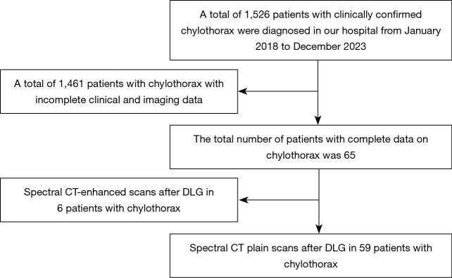
Methods: From January 2018 to December 2023, we retrospectively recruited 59 patients with clinically confirmed diagnosis of chylothorax, and all patients underwent spectral CT after DLG scanning to observe the abnormalities of lymphatic vessels and the CT findings of other thoracic abnormalities, including abnormal contrast deposition, abnormal changes in the lungs, pleura, thoracic cavity, and mediastinum.
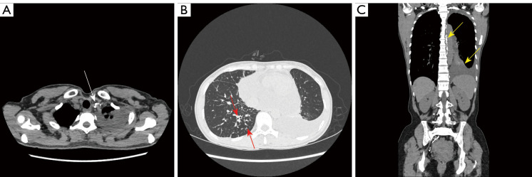
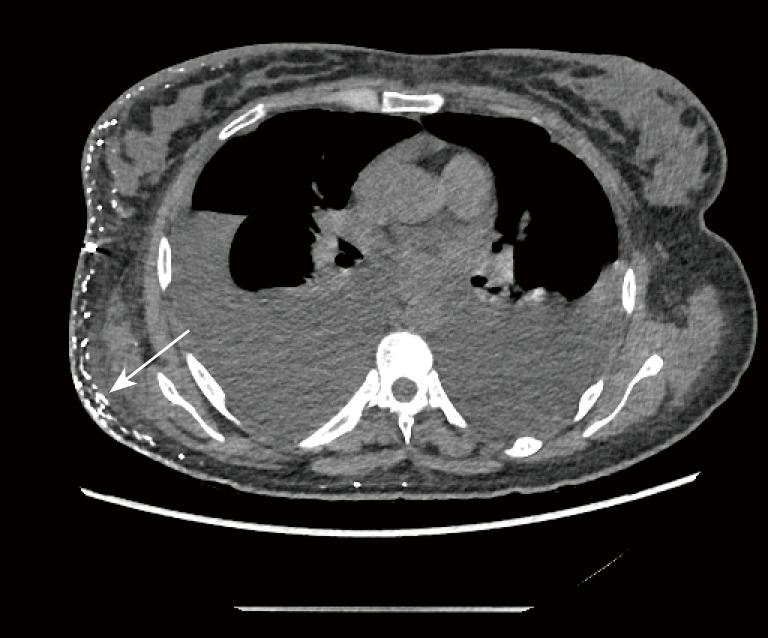
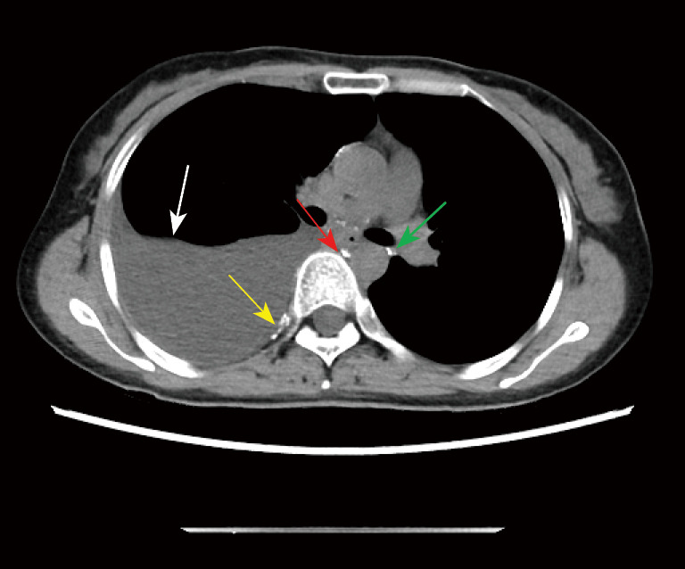
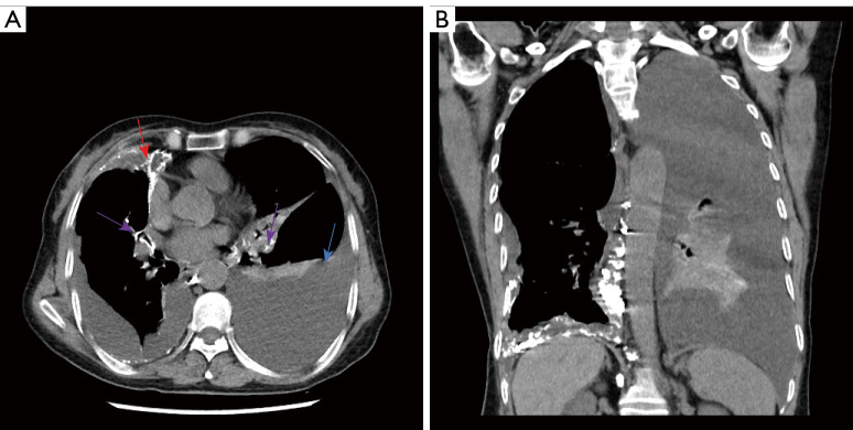
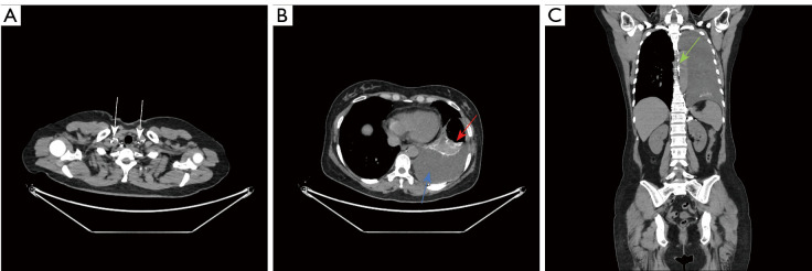
Results: In all the 59 patients with chylothorax, abnormal contrast deposition was seen on spectral CT plain scan after DLG, and could be seen simultaneously in different areas of the superior mediastinum, the mediastinum and both sides of the mediastinum. In terms of anatomical sites, the location of abnormal contrast agent deposition was most common in the left venous angle. And multiple abnormalities in the lungs, the pleura, and the mediastinum were seen.
Conclusions: Spectral CT plain scan after DLG is valuable for the diagnosis of chylothorax. Abnormal distribution of contrast in the lungs, lymphatic ducts and lymphatic trunks in spectral CT plain scan after DLG is an important manifestation of chylothorax, suggesting abnormal distribution and severity of the thoracic organs, lymphatic ducts and lymphatic trunks, and its imaging findings provide a basis for the clinical diagnosis and treatment of the disease.
Keywords: Chylothorax; X-ray computed; lymphography; tomography.
Copyright © 2025 AME Publishing Company. All rights reserved.
Conflict of interest statement
Conflicts of Interest: All authors have completed the ICMJE uniform disclosure form (available at https://qims.amegroups.com/article/view/10.21037/qims-24-1472/coif). The authors have no conflicts of interest to declare.
Figures
Similar articles
-
Computed tomography lymphangiography findings in 27 cases of lymphangioleiomyomatosis.Acta Radiol. 2017 Nov;58(11):1342-1348. doi: 10.1177/0284185116688381. Epub 2017 Jan 29. Acta Radiol. 2017. PMID: 28132530
-
Lymphatic plastic bronchitis: a study based on CT and MR lymphangiography.BMC Med Imaging. 2024 Dec 23;24(1):348. doi: 10.1186/s12880-024-01504-0. BMC Med Imaging. 2024. PMID: 39716172 Free PMC article.
-
Diagnosis and localization of laceration of the thoracic duct: usefulness of lymphangiography and CT.AJR Am J Roentgenol. 1991 Oct;157(4):703-5. doi: 10.2214/ajr.157.4.1892021. AJR Am J Roentgenol. 1991. PMID: 1892021
-
Interventional Treatment of Pulmonary Lymphatic Anomalies.Tech Vasc Interv Radiol. 2016 Dec;19(4):299-304. doi: 10.1053/j.tvir.2016.10.005. Epub 2016 Oct 8. Tech Vasc Interv Radiol. 2016. PMID: 27993326 Review.
-
Abnormal pulmonary lymphatic flow in patients with paediatric pulmonary lymphatic disorders: Diagnosis and treatment.Paediatr Respir Rev. 2020 Nov;36:15-24. doi: 10.1016/j.prrv.2020.07.001. Epub 2020 Jul 10. Paediatr Respir Rev. 2020. PMID: 32792289 Review.
References
-
- Yazicioglu A, Yazici U, Aydin E, Karaoglanoglu N. A strange bullet which caused chylomediastinum and chyloptysis. Thorac Cardiovasc Surg 2014;62:372-4. - PubMed
LinkOut - more resources
Full Text Sources