

Bibliometric analysis of the application of artificial intelligence in orthopedic imaging
- PMID: 40384704
- PMCID: PMC12084720
- DOI: 10.21037/qims-24-1384
Bibliometric analysis of the application of artificial intelligence in orthopedic imaging
Abstract
Background: With the development of artificial intelligence (AI) and the increasing significance of imaging in orthopedics, the application of AI in the field of orthopedic imaging is becoming increasingly extensive. Previous studies show that the application of AI-based orthopedic imaging may break the traditional model of the field. As a result, relevant research has received attention, and numerous articles have been published. Through bibliometric analysis, this study summarized the knowledge structure of AI-based orthopedic imaging and explored its potential research trends and focal points.
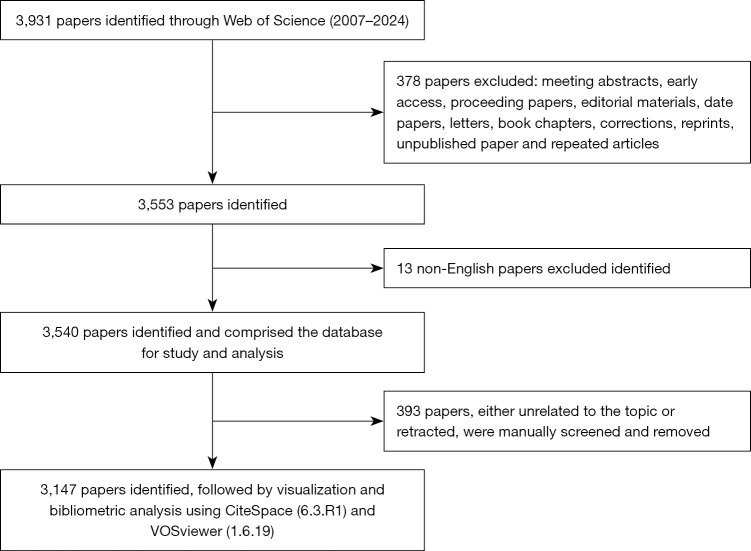
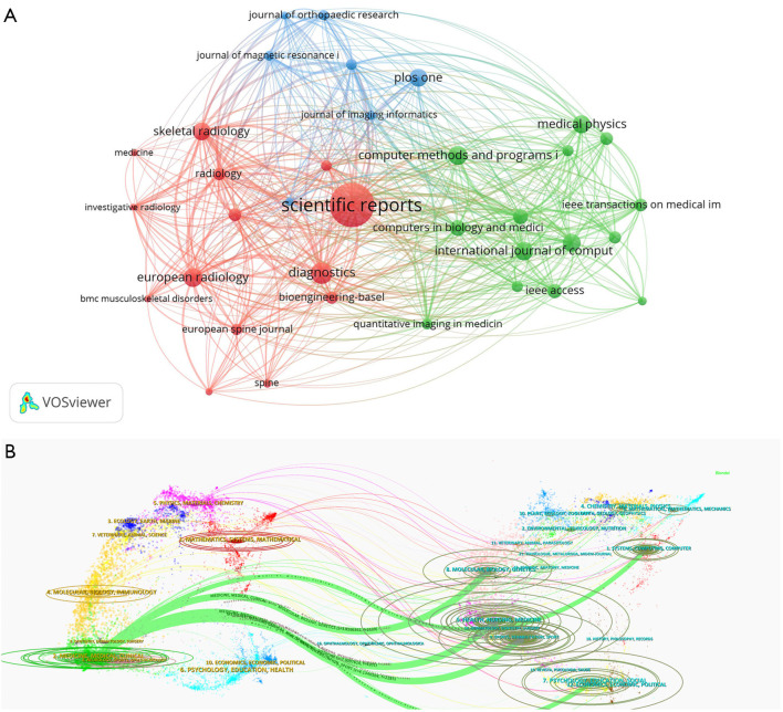
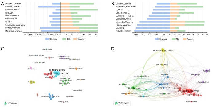
Methods: In this study, literature on AI in the field of orthopedic imaging available in the Web of Science Core Collection (WoSCC) database from 1 January 2007 to 31 December 2024 was analyzed. In order to identify the main research topics and generate visual charts of countries, institutions, authors, and keyword networks, the search results were imported into VOSviewer and CiteSpace.
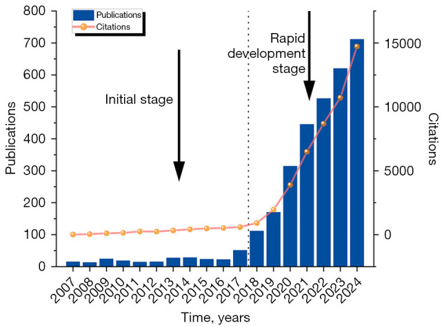
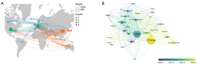
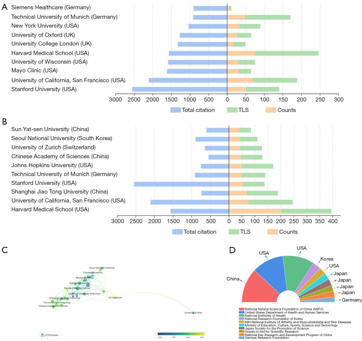
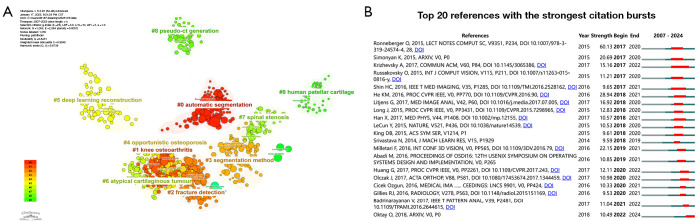
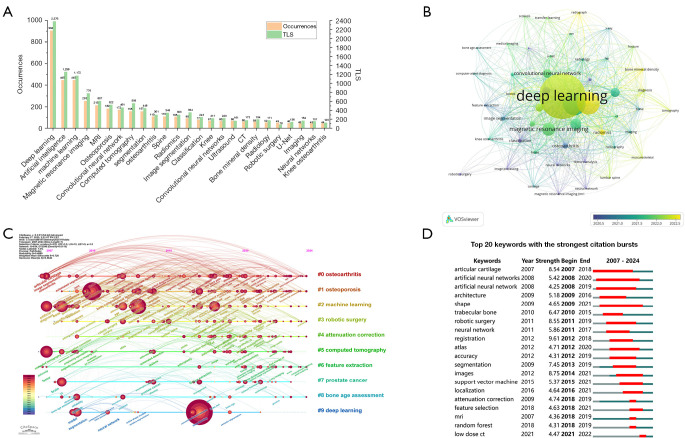
Results: A total of 3,147 publications were analyzed, revealing a rapid increase in AI research in orthopedic imaging since 2007, with over 90% of studies published after 2017. The United States (US) and China dominate this field, with the US leading in citations and academic influence, and China demonstrating significant growth in productivity. Institutional analysis highlighted Harvard University and Stanford University as key contributors, reflecting their strong academic influence. Keyword analysis identified three main research focuses: (I) advancements in algorithm development, particularly deep learning (DL) methods such as convolutional neural networks (CNNs); (II) applications in orthopedic disease imaging, including osteoarthritis, osteoporosis, and total knee arthroplasty; and (III) innovations in multimodal fusion and three-dimensional (3D) imaging techniques. Emerging trends emphasize integrating imaging data with clinical biomarkers to improve diagnostic accuracy and therapeutic decision-making. These findings provide a comprehensive overview of AI's role in orthopedic imaging, emphasizing areas of high impact and potential future directions for research.
Conclusions: The research on the application of AI in orthopedic imaging is a hot topic and indicates broad research prospects in the future. However, this study suggests that research teams should strengthen collaboration, especially international cooperation. Based on comprehensive analysis, the development of DL algorithms (especially CNNs), the use of AI in processing image data related to orthopedic diseases (segmentation, classification, and feature map extraction), and the expansion of AI imaging applications in different diseases are expected to become hotspots in future research on the application of AI in orthopedic imaging.
Keywords: Bibliometric analysis; artificial intelligence (AI); image; orthopedic.
Copyright © 2025 AME Publishing Company. All rights reserved.
Conflict of interest statement
Conflicts of Interest: All authors have completed the ICMJE uniform disclosure form (available at https://qims.amegroups.com/article/view/10.21037/qims-24-1384/coif). The authors have no conflicts of interest to declare.
Figures
Similar articles
-
Application of artificial intelligence in Alzheimer's disease: a bibliometric analysis.Front Neurosci. 2025 Feb 14;19:1511350. doi: 10.3389/fnins.2025.1511350. eCollection 2025. Front Neurosci. 2025. PMID: 40027465 Free PMC article. Review.
-
Application of artificial intelligence in palliative care: a bibliometric analysis of research hotspots and trends.Front Med (Lausanne). 2025 May 21;12:1597195. doi: 10.3389/fmed.2025.1597195. eCollection 2025. Front Med (Lausanne). 2025. PMID: 40470051 Free PMC article.
-
Artificial intelligence-assisted multimodal imaging for the clinical applications of breast cancer: a bibliometric analysis.Discov Oncol. 2025 Apr 16;16(1):537. doi: 10.1007/s12672-025-02329-1. Discov Oncol. 2025. PMID: 40237900 Free PMC article.
-
Research Trends in the Application of Artificial Intelligence in Oncology: A Bibliometric and Network Visualization Study.Front Biosci (Landmark Ed). 2022 Aug 31;27(9):254. doi: 10.31083/j.fbl2709254. Front Biosci (Landmark Ed). 2022. PMID: 36224012
-
Global output of clinical application research on artificial intelligence in the past decade: a scientometric study and science mapping.Syst Rev. 2025 Mar 15;14(1):62. doi: 10.1186/s13643-025-02779-2. Syst Rev. 2025. PMID: 40089747 Free PMC article.
References
-
- Maksymowych WP, Lambert RG, Østergaard M, Baraliakos X. Response to: ‘Correspondence on ‘MRI lesions in the sacroiliac joints of patients with spondyloarthritis: an update of definitions and validation by the ASAS MRI working group’’ by Jibri et al. Ann Rheum Dis 2023;82:e122. 10.1136/annrheumdis-2021-220078 - DOI - PubMed
LinkOut - more resources
Full Text Sources
Research Materials