. 2025 Mar 28:31:85-97.
eCollection 2025.

DNA methyltransferase 1 regulates epithelial cell functions in corneal and eyelid development

Affiliations

DNA methyltransferase 1 regulates epithelial cell functions in corneal and eyelid development

Antonius Christianto et al. Mol Vis. .

Abstract

Purpose: DNA methyltransferase 1 (DNMT1) is a crucial enzyme for the development of the retina and lens in the eye, but its roles in the cornea and eyelids are yet to be investigated.

Methods: Ocular surface epithelium (OSE)-specific Dnmt1 knockout mice, denoted as Dnmt1ΔOSE , were generated. Prenatal eye tissues were characterized by hematoxylin and eosin staining; DNMT1 expression, DNA methylation, epithelial differentiation and cell-cell junctions were determined by immunohistochemistry; proliferation was assessed by 5-ethynyl 2´-deoxyuridin labeling and apoptosis evaluated by terminal deoxynucleotidyl transferase dUTP nick-end labeling assay. Keratinocytes derived from Dnmt1F/F mice were infected with adenoviruses carrying either green fluorescent protein or Cre recombinase to obtain wild-type and Dnmt1-deficient cells. In these cells, Dnmt1 expression and epithelial terminal differentiation were evaluated by real-time PCR and/or western blotting; adherence junction and apoptosis were assessed by immunohistochemistry; proliferation was determined by 5-ethynyl 2´-deoxyuridin labeling; transcription factor activities were determined by luciferase reporter assays.

Results: The abundant DNMT1 expression and cytosine methylation (5meC) detected in the ocular surface epithelia of wild-type embryos were largely diminished in that of Dnmt1ΔOSE embryos. Besides lens degeneration, the Dnmt1ΔOSE fetuses had severe abnormalities of the cornea and eyelids. The surface epithelial cells and stromal keratocytes in the knockout corneas were distorted and the eyelids failed to fuse in the knockout embryos, resulting in an eye-open-at-birth phenotype. At the cellular level, DNMT1-deficient OSE had normal proliferation but increased apoptosis and aberrant cell junctions. In addition, the knockout corneal epithelia failed to express corneal-specific keratin 12, and the knockout eyelid epithelia had increased expression of keratin 10, indicating accelerated terminal differentiation. In vitro studies validated that DNMT1 was required for epithelial cell survival, terminal differentiation and cell junctions, and further identified signaling pathways aberrantly activated by its ablation.

Conclusion: DNMT1 maintains survival and differentiation of corneal and eyelid epithelium for the development of the eye.


Figures

Figure 1
Figure 1
Defective DNA methyltransferase 1 expression and DNA methylation in the ocular surface epithelium of Dnmt1ΔOSE embryos. Wild-type and Dnmt1ΔOSE E15.5 embryonic eye sections were examined by immunostaining for (A) DNMT1 and (B) 5meC. Dashed lines indicate the epithelial basement membrane. Top panels, low magnification images; middle and lower panels, higher magnifications of selected areas of (a) the eyelid and (b) the cornea, respectively. Arrowheads point to the eyelid epithelium in (a) and the corneal epithelium in (b), the asterisk marks the lens epithelium in (b). EL, eyelid; CO, cornea; RE, retina; LN, lens. Images are representative of at least three mice/genotypes. Scale bars: 50 µm.
Figure 2
Figure 2
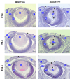
Abnormalities of the eyelids, cornea and lens in Dnmt1ΔOSE eyes. Wild-type and Dnmt1ΔOSE eye sections at the embryonic days indicated were subjected to hematoxylin and eosin staining. Arrowheads point to the eyelid tip leading edge. $ indicates corneal edema, * indicates the degenerated lens and # indicates the cell clumps above and below the degenerative lens in the Dnmt1ΔOSE eyes. EL, eyelid; CO, cornea; RE, retina; LN, lens. Images are representative of at least three mice/genotypes. Scale bars: 50 µm.
Figure 3
Figure 3
DNA methyltransferase 1 maintains cornea integrity and differentiation. A: High magnification of hematoxylin and eosin-stained images of the corneas of wild-type and Dnmt1ΔOSE E16.5 embryos. The dashed lines indicate corneal stroma junctions with the epithelium and endothelium. Arrows point to abnormal-shaped epithelial cells, asterisks show basal cells detached from the basement membrane and arrowheads point to the abnormal stromal cells in the Dnmt1ΔOSE cornea. EP, epithelium, ST, stroma, ED, endothelium. B: Measurement of the stroma thickness (left) and quantification of keratocytes (right) in wild-type and Dnmt1ΔOSE corneas. Values are mean ± s.e.m. of three embryos/genotype. ***p < 0.001 (two-tailed Student t test). Immunohistochemistry of the corneas epithelium in E15.5 embryos using (C) anti-E-cadherin (E-Cad), (D) anti-ZO-1, (E) anti-K14 and (F) anti-K12 at low (upper panels) and high (lower panels) magnifications of the selected areas (a, lower panels). Dotted lines indicate corneal epithelial basement membranes. White arrows indicate epithelial cell detachment from the basement membrane. Images are representative of at least three mice/genotypes. Scale bars: 10 µm for (A), (C) and (D). 30 µm for (E) and (F).
Figure 4
Figure 4
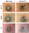
Eye-open phenotypes of Dnmt1ΔOSE mice. Photographs of wild-type and Dnmt1ΔOSE eyes at different developmental stages. Wild-type pups showed eyelid fusion but Dnmt1ΔOSE pups displayed eye-open phenotypes (arrows). Images are representative of at least three mice/genotypes. Scale bar: 1 mm.
Figure 5
Figure 5
DNA methyltransferase 1 ablation disrupts eyelid morphology and differentiation. A: Wild-type and Dnmt1ΔOSE E15.5 embryonic eye sections were subjected to Hematoxylin and eosin staining, and eyelid tip images were captured at lower (upper panels) and higher magnifications of the selected areas (a, lower panels). Arrowheads point to the epithelial protrusion of the eyelid tip, asterisks mark eyelid epithelial folding and arrows indicate cells displaying condensed nuclei in the Dnmt1ΔOSE eyelids. B: Immunostaining using anti-E-Cad and anti-ZO-1, as indicated. Arrows point to abnormal ZO-1 localization and reduced E-Cad in the Dnmt1ΔOSE eyelids. C: Wild-type and Dnmt1ΔOSE embryonic eyelids at embryonic days indicated were subjected to immunostaining with anti-K10 for epithelial terminal differentiation. Images were taken at low magnification (upper panels) and high magnification of selected areas (a, lower panels). Arrowheads point to different K10 expression between wild-type and Dnmt1ΔOSE eyelid tips. Dashed lines indicate the epithelial basement membrane. Images are representative of at least three mice/genotypes. Scale bars: 10 µm for (A) and (B). Scale bars: 30 µm for (C). EP, epithelium; DE, dermis.
Figure 6
Figure 6
Apoptosis and proliferation in wild-type and Dnmt1ΔOSE embryos. A: Representative images of the terminal deoxynucleotidyl transferase dUTP nick-end labeling assay showing apoptotic cells (green, arrows) in different regions of the ocular surface epithelium. B: Illustration of the epithelial regions for the eyelids (red), cornea (green) and lens (yellow) used for quantification. Quantification of (C) apoptotic cells and (D) proliferating cells in the different regions of the ocular surface epithelium in E14.5–E16.5 embryos. EL, Eyelid; CO, Cornea; LN, Lens. Values are mean ± s.e.m. of three embryos/genotype. *p < 0.05, **p < 0.01 (two-tailed Student t test).
Figure 7
Figure 7
The role of DNMT1 in cultured keratinocytes. Dnmt1F/F keratinocytes infected with Ad-GFP or Ad-Cre were examined for (A) Dnmt1 mRNA and (C) K14, K1 and K10 mRNA, normalized by mRNA of Gapdh. (B) DNMT1 protein normalized by β-actin. D: The percentage of apoptosis and proliferation quantified by immunolabelling with anti-Cas-3 and anti-EdU assays, respectively. E: Immunofluorescent staining using anti-E-cadherin (E-Cad). Arrowhead points to Ad-GFP/Ad-GFP-Cre–infected cells. Scale bar: 30 µm. F: Dnmt1F/F reporter stable cells were infected with Ad-GFP or Ad-Cre, and luciferase activities were normalized by total protein. Values are mean ± s.e.m. of three wells/adenovirus. *p < 0.05, ***p < 0.001 (two-tailed Student t test).

Similar articles

References

    1. Jin B, Li Y, Robertson KD. DNA methylation: superior or subordinate in the epigenetic hierarchy? Genes Cancer. 2011;2:607–17. - PMC - PubMed
    1. Bird A. DNA methylation patterns and epigenetic memory. Genes Dev. 2002;16:6–21. - PubMed
    1. Bernstein BE, Kamal M, Lindblad-Toh K, Bekiranov S, Bailey DK, Huebert DJ, McMahon S, Karlsson EK, Kulbokas EJ, 3rd, Gingeras TR, Schreiber SL, Lander ES. Genomic maps and comparative analysis of histone modifications in human and mouse. Cell. 2005;120:169–81. - PubMed
    1. Kikuchi A, Onoda H, Yamaguchi K, Kori S, Matsuzawa S, Chiba Y, Tanimoto S, Yoshimi S, Sato H, Yamagata A, Shirouzu M, Adachi N, Sharif J, Koseki H, Nishiyama A, Nakanishi M, Defossez PA, Arita K. Structural basis for activation of DNMT1. Nat Commun. 2022;13:7130. - PMC - PubMed
    1. Egger G, Jeong S, Escobar SG, Cortez CC, Li TW, Saito Y, Yoo CB, Jones PA, Liang G. Identification of DNMT1 (DNA methyltransferase 1) hypomorphs in somatic knockouts suggests an essential role for DNMT1 in cell survival. Proc Natl Acad Sci U S A. 2006;103:14080–5. - PMC - PubMed

MeSH terms

Substances

LinkOut - more resources