. 2024 Apr 19;5(1):33-49.
doi: 10.5194/mr-5-33-2024. eCollection 2024.

Deuteration of proteins boosted by cell lysates: high-resolution amide and H α magic-angle-spinning (MAS) NMR without the reprotonation bottleneck

Affiliations

Deuteration of proteins boosted by cell lysates: high-resolution amide and H α magic-angle-spinning (MAS) NMR without the reprotonation bottleneck

Federico Napoli et al. Magn Reson (Gott). .

Abstract

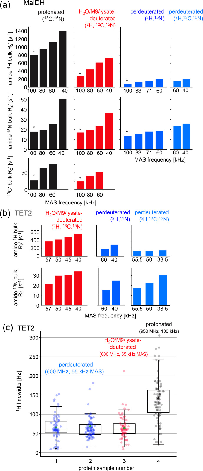
Amide-proton-detected magic-angle-spinning NMR of deuterated proteins has become a main technique in NMR-based structural biology. In standard deuteration protocols that rely on D 2 O-based culture media, non-exchangeable amide sites remain deuterated, making these sites unobservable. Here we demonstrate that proteins produced with a H 2 O-based culture medium doped with deuterated cell lysate allow scientists to overcome this "reprotonation bottleneck" while retaining a high level of deuteration (ca. 80 %) and narrow linewidths. We quantified coherence lifetimes of several proteins prepared with this labeling pattern over a range of magic-angle-spinning (MAS) frequencies (40-100 kHz). We demonstrate that under commonly used conditions (50-60 kHz MAS), the amide 1 H linewidths with our labeling approach are comparable to those of perdeuterated proteins and better than those of protonated samples at 100 kHz. For three proteins in the 33-50 kDa size range, many previously unobserved amides become visible. We report how to prepare the deuterated cell lysate for our approach from fractions of perdeuterated cultures which are usually discarded, and we show that such media can be used identically to commercial media. The residual protonation of H α sites allows for well-resolved H α -detected spectra and H α resonance assignment, exemplified by the de novo assignment of 168 H α sites in a 39 kDa protein. The approach based on this H 2 O/cell-lysate deuteration and MAS frequencies compatible with 1.3 or 1.9 mm rotors presents a strong sensitivity benefit over 0.7 mm 100 kHz MAS experiments.


Conflict of interest statement

At least one of the (co-)authors is a member of the editorial board of Magnetic Resonance. The peer-review process was guided by an independent editor, and the authors also have no other competing interests to declare.

Figures

Figure 1
Figure 1
Deuteration using H 2 O-based medium doped with algal lysate. (a) Deuteration level of Ignicoccus islandicus MalDH (33.55 kDa) expressed in H 2 O with either 2 H, 13 C glucose only (2 g per liter of culture) and 15 NH 4 or with additional use of 2 H, 13 C, 15 N algal extract (ISOGRO®) at three different concentrations (1, 2, 4 g per liter of culture). The reported molecular-weight values are from intact mass spectrometry. The dashed red line indicates the theoretical molecular weight of fully deuterated MalDH (assuming all exchangeable hydrogens are 1 H). (b, c) Residual protonation level for aliphatic sites, determined by solution-state NMR of a sample of ubiquitin produced in H 2 O-based M9 medium supplemented with 2 H, 13 C, 15 N algal extract (2 g L -1 ). Amino acid types missing in this plot are either not present in ubiquitin, excluding the possibility for quantification (Trp), or are not visible or unresolved (Met, His, Arg).
Figure 2
Figure 2
Comparison of the amino acid composition in a home-made bacterial extract with the one in ISOGRO®. (a) The amino acid composition of ISOGRO®, as provided by the manufacturer, is shown as grey bars; the values represent the percentage (weight) of each amino acid in the powder. Data of the home-made preparation from bacterial perdeuterated cultures are shown in blue and red. The preparation made from the soluble-protein fraction, i.e., contaminant proteins from a protein purification, is shown in red. The preparation made from the insoluble fraction after cell lysis is shown in blue. Asn and Gln are indistinguishable from Asp and Glu with this method. Cys and Trp are destroyed during the processing steps of the analysis. (b) 1D 1 H spectra of ubiquitin deuterated in a H 2 O-based medium supplemented with either commercial algal lysate (black) or the home-made lysate, prepared from the contaminant proteins. The precultures have been made either in H 2 O or D 2 O, as indicated. The similarity of the spectra confirms that commercial algal lysates and bacterial lysates produce similar labeling patterns, as expected from the very similar distribution of amino acids shown in panel (a).
Figure 3
Figure 3
Investigation of coherence lifetimes and linewidths in differently labeled samples of two proteins. (a) Fitted R2 decay rate constants for four differently labeled samples of MalDH as indicated. The decay curves for representative experiments, indicated with an asterisk, are shown in Fig. S1. All data were recorded at a B 0 field strength corresponding to 700 MHz 1 H Larmor frequency in a 0.7 mm probe. (b) Similar data for the TET2 protein. (c)  1 H linewidths of TET2 in deuterated samples produced in D 2 O-based M9 medium (blue), produced in a H 2 O-based M9 medium doped with 2 g L -1 deuterated algal extract (red) or fully protonated (black). Spectra are shown in Fig. S2.
Figure 4
Figure 4
Deuteration in H 2 O enhanced by amino acid mixtures from cell lysates allows for the detection of non-exchangeable amide hydrogens. Overlay of (a) hNH, (b) hCANH, (c) hCONH TET2 spectra, (d) H–N, and (e) CA–N projections of the hCANH MalDH spectra, as well as (f) hCANH pb6 spectra. (For the latter, the in-house bacterial cell lysate was used.) Spectra of samples deuterated by the H 2 O/M9/lysate approach are shown in red, while spectra from perdeuterated samples are shown in blue. All samples were 13 C and 15 N labeled. Peaks that appear to only be present in the perdeuterated sample spectra can be explained by (i) different apparent frequencies of aliased peaks (since the spectral widths and carriers are not identical in the overlaid spectra), (ii) chemical shift perturbations due to small sample-temperature differences, and (iii) different signal-to-noise ratios of individual peaks.
Figure 5
Figure 5
Newly assigned amide hydrogens in TET2 deuterated in H 2 O shown on its monomeric structure. Amide 1 H frequencies that were previously inaccessible with perdeuterated samples and that were assigned with the H 2 O/M9/lysate deuteration are represented as spheres on the protein structure. Blue spheres correspond to atoms that could be assigned from the spectra of the H 2 O/M9/lysate-deuterated sample (600 MHz 1 H Larmor frequency, 55 kHz MAS), while cyan spheres are for atoms that only showed signal in the spectra of the fully protonated sample (950 MHz 1 H Larmor frequency, 100 kHz MAS). Parts for which the backbone is shown in white regions correspond to those for which heavy-atom assignments have previously been reported , while black regions indicate parts with missing heavy-atom assignment. In those parts we did not attempt to get new amide assignments, as the de novo assignment of the backbone would likely require more 3D data sets. Taken together, these data highlight the possibility of detecting amide hydrogens of water-inaccessible regions in the protein. Note that the protein forms a dodecamer (Fig. a), and for better visibility only the monomeric subunit is shown.
Figure 6
Figure 6
Protein deuteration in H 2 O allows assignment of H α hydrogens. (a) Example strips from 4D HACANH and (b) 3D hNCAHA and hCANH correlation experiments exploited for the assignment of H α hydrogens. (c) Overlay of the H α region of the hCH spectra for the fully protonated (grey) and deuterated in H 2 O (red) samples. The same color scheme applies to panels (a) and (b). (d) Newly assigned H α hydrogens are represented as spheres on the protein structure. Green spheres correspond to atoms that could be assigned from the spectra of the sample deuterated in H 2 O, while orange spheres are for atoms that only showed signal in the fully protonated sample's spectra. Black regions indicate missing H α assignment. (e) Sequence-based representation of TET2 resonance assignments. Newly assigned amide and H α hydrogens are shown in blue and green, respectively. Lost assignments correspond to sites for which the automated-assignment software FLYA did not converge after adding the peak list from the H 2 O/M9/lysate-deuterated sample to the ones used previously for automatic assignment . Protein deuteration in H 2 O allows assignment of H α hydrogens.

Similar articles

Cited by

References

    1. Agarwal V, Penzel S, Szekely K, Cadalbert R, Testori E, Oss A, Past J, Samoson A, Ernst M, Böckmann A, Meier B H. De novo 3D structure determination from sub-milligram protein samples by solid-state 100 kHz MAS NMR spectroscopy. Angew Chem Int Edit. 2014;53:12253–12256. - PubMed
    1. Anderson E. Growth requirements of virus-resistant mutants of Escherichia coli strain “B”. P Natl Acad Sci USA. 1946;32:120–128. - PMC - PubMed
    1. Andreas LB, Stanek J, Le Marchand T, Bertarello A, Paepe DC-D, Lalli D, Krejčíková M, Doyen C, Öster C, Knott B, Wegner S, Engelke F, Felli IC, Pierattelli R, Dixon NE, Emsley L, Herrmann T, Pintacuda G. Protein residue linking in a single spectrum for magic-angle spinning NMR assignment. J Biomol NMR. 2015;62:253–261. - PubMed
    1. Andreas LB, Jaudzems K, Stanek J, Lalli D, Bertarello A, Le Marchand T, Cala-De Paepe D, Kotelovica S, Akopjana I, Knott B, Wegner S, Engelke F, Lesage A, Emsley L, Tars K, Herrmann T, Pintacuda G. Structure of fully protonated proteins by proton-detected magic-angle spinning NMR. P Natl Acad Sci USA. 2016;113:9187–9192. - PMC - PubMed
    1. Arnaud C-A, Effantin G, Vivès C, Engilberge S, Bacia M, Boulanger P, Girard E, Schoehn G, Breyton C. Bacteriophage T5 tail tube structure suggests a trigger mechanism for Siphoviridae DNA ejection. Nat Commun. 2017;8:1953. doi: 10.1038/s41467-017-02049-3. - DOI - PMC - PubMed

LinkOut - more resources