Review
. 2025 May 2:12:1492275.
doi: 10.3389/frobt.2025.1492275. eCollection 2025.

Exoskeletons for the rehabilitation of temporomandibular disorders: a comprehensive review

Affiliations
Review

Exoskeletons for the rehabilitation of temporomandibular disorders: a comprehensive review

Paul-Otto Müller et al. Front Robot AI. .

Abstract

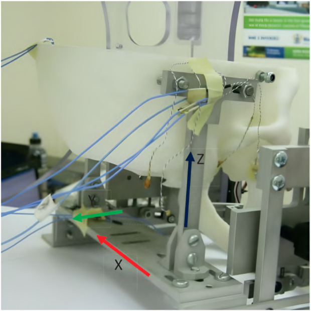
Despite the many technological advancements in exoskeletons for the rehabilitation of lower or upper limbs, there has been limited exploration of their application in treating temporomandibular disorders, a set of musculoskeletal and neuromuscular conditions affecting the masticatory system. By collecting data, implementing assisting and resisting training routines, and encouraging active patient engagement, exoskeletons could provide controlled and individualized exercise with flexibility in time and location to aid in the recovery or improvement of jaw mobility and function. Thus, they might offer a valuable alternative or complement to conservative physiotherapy. In this context, the review aims to draw attention to rehabilitating temporomandibular disorders with the help of exoskeletons by looking at the advantages and opportunities these devices potentially provide. After stating the requirements and resulting scientific challenges in various fields and discussing the state of the art, existing research gaps and deficiencies will be discussed, highlighting areas where further research and development is needed.

Keywords: TMD; exoskeletons; physical therapy; rehabilitation; review; robotics; temporomandibular disorders.


Conflict of interest statement

The authors declare that the research was conducted in the absence of any commercial or financial relationships that could be construed as a potential conflict of interest.

Figures

FIGURE 1
FIGURE 1
An illustration of the jaw anatomy, highlighting key structures involved in jaw motion and their relevance to the masticatory system. The image displays bony structures in white, including the mandible (lower jaw), maxilla (upper jaw), and hyoid bone, which provide the structural framework for the masticatory system. In red, a selection of muscles responsible for opening and closing the jaw, such as the digastric, masseter, and parts of the pterygoid muscles, are shown. These muscles play critical roles in enabling complex jaw movements such as elevation, depression, protrusion, and lateral excursion. The articular disc, depicted in blue, is positioned between the mandibular condyle and the mandibular fossa of the temporal bone. This disc facilitates smooth articulation by absorbing shear and compressive forces during jaw motion. The illustration excludes ligaments and other soft tissues, such as the temporomandibular joint capsule, which also contribute to joint stability and motion control. This depiction serves as a simplified anatomical reference for understanding the mechanical and biomechanical interactions involved in jaw rehabilitation, particularly in the context of temporomandibular disorders. Information from Okeson was taken as a reference to create the image (Okeson, 2019).
FIGURE 2
FIGURE 2
PRISMA flow chart of the literature search and study selection process (Page et al., 2021). The literature search, conducted in July 2024, included six different databases, two of which were grey literature databases, resulting in a total of 4,275 records. After removing 546 duplicates by using the JabRef bibliography management tool (Kopp et al., 2023), 3,729 unique records were screened for relevance based on their titles and abstracts. Of these, 23 full-text articles were assessed for eligibility, with 17 articles excluded for various reasons, including the unavailability of an English text, lack of wearability and reversibility in the rehabilitation method, and invasiveness. Ultimately, six publications were deemed eligible and included in the final literature review.
FIGURE 3
FIGURE 3
Two versions of the jaw exoskeleton developed by Wang et al. (a) The first version comprises a rigid four-bar linkage system attached to a helmet, driven by a DC motor placed on top of the helmet and connected via a belt to the linkage system. The chin is placed in between two cushioned bars to transmit the forces. The links are adjustable to accommodate different patients and trajectories. A model of a human mandible can be seen in between the chin holder bars. ©2010 IEEE. Reprinted with permission. (b) The second version is similar in design but features a more compact structure. Two DC motors are placed on the sides of the helmet and are connected to the linkage system via gears. The chin holder has a more complex design, and hidden springs are added to introduce passive compliance to the system. A human lower jaw is depicted in red. The material used for the linkage system in both designs is aluminum. Reprinted with permission by the author. (a) The first device version by Wang (2010). (b) The second device version Wang (2014).
FIGURE 4
FIGURE 4
A 3D-printed model of the skull and jaw to evaluate the jaw exoskeleton concept. The skull is fixed to a base plate, and the exoskeleton is attached to the freely moving jaw. Sensors are placed inside the joints measuring forces in a range of 15 N–60 N during the interaction. The condyles and incisor points are tracked by an electromagnetic-based recording device (Wang, 2014). Reprinted with permission by the author.
FIGURE 5
FIGURE 5
An illustration of the jaw exoskeleton concept by Evans et al. (2016). (a) The image shows the device with the shoulder mount, the head brace, and the mouthpiece linked through a linear slide to a bracket attached to an actuator system. ©2016 IEEE. Reprinted with permission. (b) The device is mounted on the patient’s shoulder and provides assistance in the vertical direction. The mouthpiece can passively compensate for the mandible’s translational motions through a linear slide. A brace fixes the head in place. The processing unit is worn on the back of the device and patient, connected to the motor by a cable shown in turquoise. ©2016 IEEE. Reprinted with permission.
FIGURE 6
FIGURE 6
The drive mechanism, mouthpiece, and control system of the jaw exoskeleton concept by Evans et al. (2016). (Top left) The drive mechanism redirects the torque via a gear system from the vertically placed DC motor over the bracket to the mouthpiece. Only vertical assistance can be provided. The bracket is rigidly attached to the gear system and includes a counterweight on the back to balance the system and reduce the load on the actuator. ©2016 IEEE. Reprinted with permission. (Top right) The mouthpiece consists of a linear slide and a strain gauge placed on the slide to measure the forces acting on the mouthpiece. A chin strap enables transmitting forces to the mandible in both vertical directions. The mandible fitting connects to the lower teeth and can be replaced. ©2016 IEEE. Reprinted with permission. (Bottom) The control system design features a hierarchical structure with a motor controller, an impedance controller, and a trajectory planner. The motor controller is responsible for the actuation, the impedance controller for the active vertical compliance, and the trajectory planner for the trajectory following tasks. The system can be cut off from power through a hand-held safety button. Adapted from Evans et al. (2016).
FIGURE 7
FIGURE 7
An overview of the exoskeleton by Kameda et al. (2021). The device implements four drive modes. In the first mode, the active part of the device is disabled, only the passive stainless steel (SS) springs act against mouth opening. In the second mode, the SMA springs are additionally heated, providing a force to follow the closing trajectory. The third mode is similar to the second but with a higher force output, actively assisting the closing motion. In the fourth mode, the device only provides assistance in the closing direction and no resistance during the opening movement. The exoskeleton itself comprises a chin cup attached to a head mount by cables and SMA and passive springs. Only forces in the closing direction can be applied. ©2021 The Japanese Society for Dental Materials and Devices. Reprinted with permission.
FIGURE 8
FIGURE 8
An illustration of the drive mechanism of the jaw exoskeleton by Kameda et al. (2021). The main actuator system is based on SMA springs, which contract when heated by an electric current. The springs expand again when cooled down. A fan is triggered by a magnetic sensor to accelerate the cooling process. Stainless steel (SS) springs are added to counteract the time delay of the SMA springs. The cup is attached to a head mount and the actuator system by cables. ©2021 The Japanese Society for Dental Materials and Devices. Reprinted with permission.
FIGURE 9
FIGURE 9
An overview of the jaw exoskeleton design and testing by Zhang et al. (2021). (a) The device consists of two pneumatic joints attached to the head and chin by an adjustable fixing ribbon. The joints are capable of providing assistance in two DoF. ©2021 IEEE. Reprinted with permission. (b) The exoskeleton was evaluated on a physical skull model, which only included the rigid bones. The mandible was actuated and the exoskeleton only provided assistance in the sagittal plane. The image shows the trajectory of two markers placed on the end-effector of the pneumatic joints. The two black lines represent the orientation of the end-effector at two distinct time points. The triangles indicate the non-actuated position of the end-effector, and the red dots indicate the actuated position. By pushing the condyles out of the fossae, a translational movement is generated leading to a wider opening of the mouth. The 2D model of the pneumatic joints shows the bellow-shaped cylinders attached to a base and an end-effector. ©2021 IEEE. Reprinted with permission. (c) The control scheme utilizes a feed-forward approach, where pressure signals are generated using an inverse kinematic model of the pneumatic joints to track a predefined trajectory. Pressure is monitored via sensors and regulated through control valves. Adapted from Zhang et al. (2021).

Similar articles

References

    1. Abboud W., Yarom N., Yahalom R., Joachim M., Reiter S., Koren O., et al. (2018). Comparison of two physiotherapy programmes for rehabilitation after temporomandibular joint arthroscopy. Int. J. Oral Maxillofac. Surg. 47, 755–761. 10.1016/j.ijom.2017.10.019 - DOI - PubMed
    1. Abdi A. H., Sagl B., Srungarapu V. P., Stavness I., Prisman E., Abolmaesumi P., et al. (2020). Characterizing motor control of mastication with soft actor-critic. Front. Hum. Neurosci. 14, 188. 10.3389/fnhum.2020.00188 - DOI - PMC - PubMed
    1. Agarwal P., Deshpande A. D. (2019). “234Exoskeletons: state-of-the-art, design challenges, and future directions,” in Human performance optimization: the science and ethics of enhancing human capabilities (Oxford University Press; ). 10.1093/oso/9780190455132.003.0011 - DOI
    1. Ariji Y., Katsumata A., Ogi N., Izumi M., Sakuma S., Iida Y., et al. (2009). An oral rehabilitation robot for massaging the masseter and temporal muscles: a preliminary report. Oral Radiol. 25, 53–59. 10.1007/s11282-009-0014-0 - DOI
    1. Ariji Y., Nakayama M., Nishiyama W., Ogi N., Sakuma S., Katsumata A., et al. (2015). Potential clinical application of masseter and temporal muscle massage treatment using an oral rehabilitation robot in temporomandibular disorder patients with myofascial pain. CRANIO® 33, 256–262. 10.1179/2151090314y.0000000030 - DOI - PubMed

LinkOut - more resources