Review
. 2024 Dec 24:10:e20240012.
doi: 10.22575/interventionalradiology.2024-0012. eCollection 2025 Mar 28.

Nodal Lymphangiography and Embolization for Postoperative Lymphatic Leakage

Affiliations
Review

Nodal Lymphangiography and Embolization for Postoperative Lymphatic Leakage

Shuji Kariya et al. Interv Radiol (Higashimatsuyama). .

Abstract

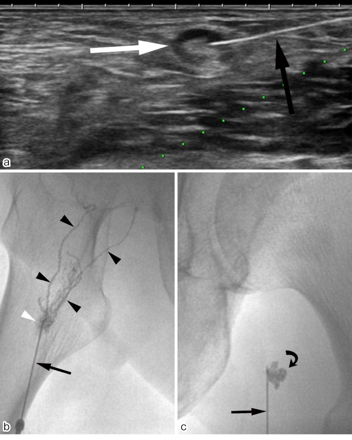
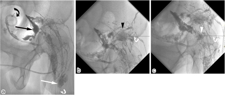
Intranodal lymphangiography has replaced conventional pedal lymphangiography and has advanced lymphatic intervention. In this method, a lymph node is punctured and Lipiodol is injected to visualize the subsequent lymphatic vessels. This has facilitated the widespread adoption of lymphatic interventional radiology due to the simplicity of the technique and the shortened examination time of the procedure, which allows easy mapping of lymphatic vessels and lymphatic fluid dynamics. With this technique, lymphatic embolization was achieved by injecting an embolic substance into the lymph nodes upstream of the lymphatic leak. Although complications associated with lymphangiography are rare, caution should be exercised due to potential complications associated with the use of Lipiodol. This study summarizes intranodal lymphangiography techniques, complications, and lymphatic embolization.

Keywords: intranodal lymphangiography; lipiodol; lymphatic embolization; lymphocele; lymphorrhea.


Conflict of interest statement

None

Figures

Figure 1.
Figure 1.
Intranodal lymphangiography. (a) A femoral lymph node (white arrow) was punctured with a 23-gauge Cathelin needle (black arrow) under ultrasound guidance. The needle was advanced until the tip reached a position between the cortex and hilum. (b) Lipiodol was injected under fluoroscopy. If the puncture is successful, fluoroscopy immediately shows granular nodules and the lymphatic vessels (black arrowheads) extending from the lymph nodes (white arrowhead). Cathelin needle (black arrow). (c) If the lymph node puncture fails, fluoroscopy shows spreading lobulated nodular pooling (curved arrow) and the lymphatic vessels are not delineated. Failure to puncture the lymph node results in a lobulated dark shadow (curved arrowheads) of leaking Lipiodol outside the node. In this case, the injection should be stopped. Cathelin needle (black arrow).
Figure 2.
Figure 2.
A 72-year-old male. A left pelvic lymphocele developed after rectal cancer surgery. A drainage catheter was placed in the lymphocele due to pressure pain. There was a daily drainage of 100 mL. After lymphatic embolization, the drainage stopped and the drainage catheter was removed 5 days after embolization. (a) Intranodal lymphangiography. The right femoral lymph node (white arrow) was punctured with a 23-gauge Cathelin needle (curved white arrow) under ultrasound guidance and Lipiodol was injected. There is leakage of Lipiodol (black arrow) into the lymphocele. Drainage catheter placed in the lymphocele (curved black arrow). (b) Lymphatic embolization. The lymph node upstream of the leak (black arrowhead) was punctured under fluoroscopy with a 23-gauge Cathelin needle (white curved arrow). Then, 2 mL of a 1:3 mixture of N-butyl cyanoacrylate and Lipiodol liquid embolic material was injected. (c) The other lymph node upstream of the leak (white arrowhead) was punctured under fluoroscopy with a 23-gauge Cathelin needle (curved white arrow). Then, 3.7 mL of a 1:3 mixture of N-butyl cyanoacrylate and Lipiodol liquid embolization material was injected.

Similar articles

References

    1. Kinmonth JB, Taylor GW, Harper RK. Lymphangiography; a technique for its clinical use in the lower limb. Br Med J. 1955; 1:940-942. - PMC - PubMed
    1. Wallace S, Jackson L, Greening RR. Clinical applications of lymphangiography. Am J Roentgenol Radium Ther Nucl Med. 1962; 88:97-109. - PubMed
    1. Cope C. Percutaneous transabdominal embolization of thoracic duct lacerations in animals. J Vasc Interv Radiol. 1996; 7:725-731. - PubMed
    1. Cope C, Salem R, Kaiser LR. Management of chylothorax by percutaneous catheterization and embolization of the thoracic duct: prospective trial. J Vasc Interv Radiol. 1999; 10:1248-1254. - PubMed
    1. Nadolski GJ, Itkin M. Feasibility of ultrasound-guided intranodal lymphangiogram for thoracic duct embolization. J Vasc Interv Radiol. 2012; 23:613-616. - PubMed