. 2025 May 2:15:1578163.
doi: 10.3389/fcimb.2025.1578163. eCollection 2025.

Mycobacterium tuberculosis modulates NUDT21-mediated alternative polyadenylation to enhance FTH1 expression in macrophages and promotes intracellular growth

Affiliations

Mycobacterium tuberculosis modulates NUDT21-mediated alternative polyadenylation to enhance FTH1 expression in macrophages and promotes intracellular growth

Xiaoqian Liu et al. Front Cell Infect Microbiol. .

Abstract

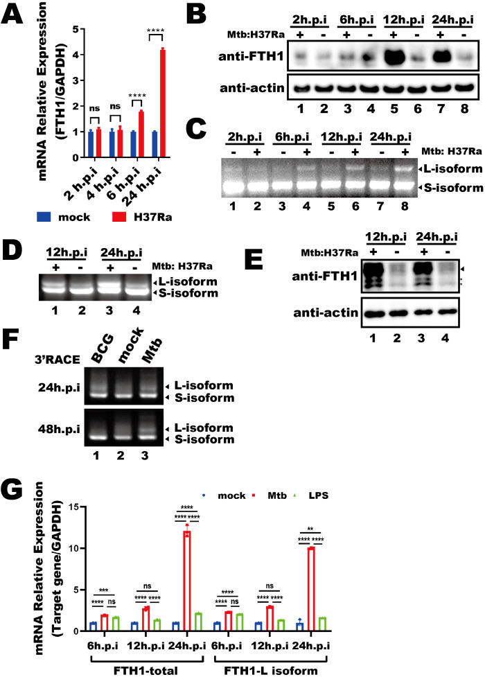
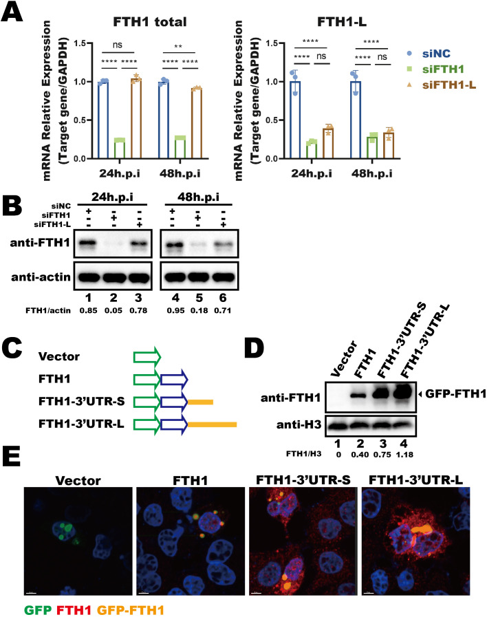
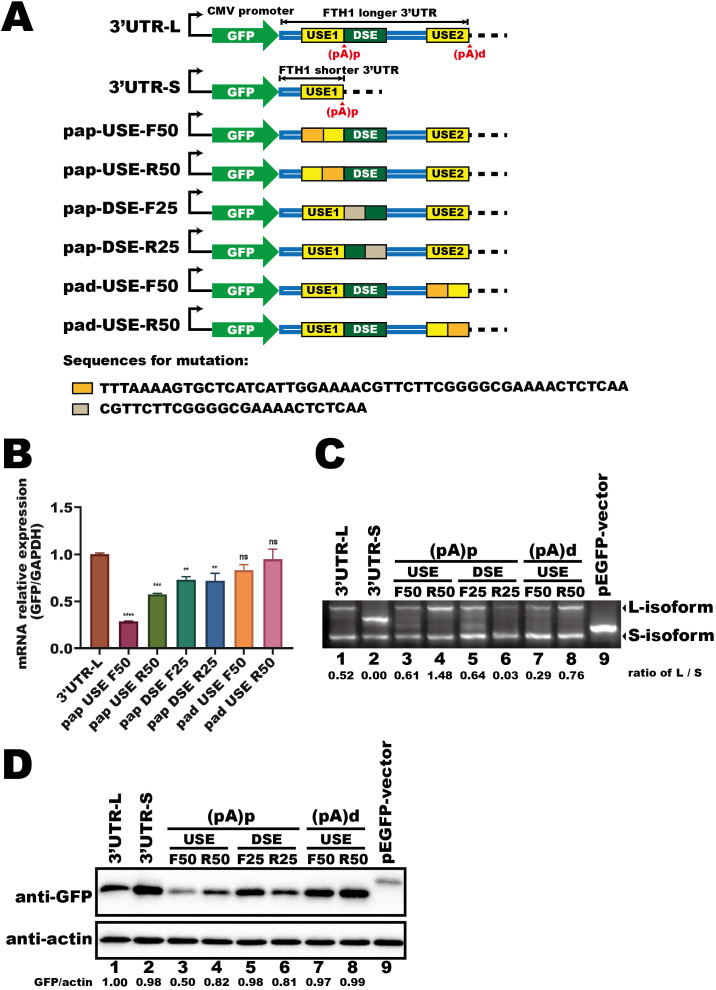
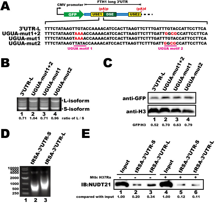
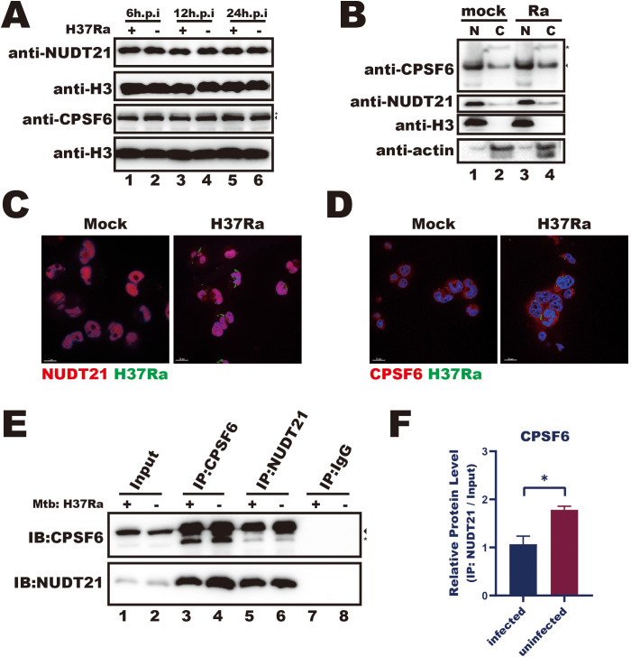
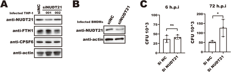
Ferritin heavy chain 1 (FTH1) is a key iron-storage protein that regulates iron availability, supports immune defense, and prevents iron-induced toxicity. During Mycobacterium tuberculosis (Mtb) infection, macrophages enhance FTH1 expression to sequestrate iron and limit Mtb growth. However, Mtb can exploit the host ferritinophagy pathway to degrade FTH1 and release iron, thereby promoting its survival. Although FTH1 plays an essential role in host-pathogen interaction during Mtb infection, its regulation remains unclear. Previous studies suggest that post-transcriptional mechanism, particularly alternative polyadenylation (APA), are critical in immune responses. We propose that APA, which determines the length of a transcript's 3'UTR, may regulate FTH1 expression during Mtb infection. Our study demonstrates that Mtb induces APA of FTH1 in macrophages, favoring the production of longer isoforms that enhance protein synthesis. Mechanistically, Mtb disrupts the interaction between NUDT21 and CPSF6, impairing NUDT21's ability to bind UGUA motifs in the FTH1 3'UTR, a key step in polyadenylation site selection. Silencing NUDT21 reduces macrophage bactericidal activity against Mtb, highlighting its role in immune defense. These findings reveal a novel Mtb-driven mechanism that enhances FTH1 expression via the NUDT21-mediated APA pathway in macrophages, suggesting that Mtb manipulates this process to promote its survival. This study provides new insights into tuberculosis pathogenesis and points to potential avenues for therapeutic exploration.

Keywords: FTH1; Mycobacterium tuberculosis; NUDT21; alternative polyadenylation; macrophage.


Conflict of interest statement

The authors declare that the research was conducted in the absence of any commercial or financial relationships that could be construed as a potential conflict of interest.

Figures

Figure 1
Figure 1
Mtb infection alters the FTH1 APA profile in macrophages. (A) qPCR analysis of FTH1 mRNA in Mtb-infected (H37Ra) or uninfected (mock) macrophages for 2-, 4-, 6-, and 24-hours post-infection. Data are normalized to GAPDH and presented as fold change relative to the control. Error bars represent the mean ± SEM from two independent experiments. Statistical significance was determined by two-way ANOVA with Sidak’s multiple comparisons test; ****P < 0.0001. (B) Western blot analysis of FTH1 protein expression in THP-1 macrophages infected with Mtb at 2, 6, 12, and 24 hours. β-actin was used as a loading control. (C) Agarose gel electrophoresis of 3′RACE products from Mtb-infected or uninfected (control) macrophages. Bands representing different FTH1 transcript isoforms are indicated by arrows. (D) 3′RACE assay of Fth1 transcripts and (E) western blot analysis of mouse FTH1 protein expression in Mtb-infected or uninfected RAW264.7 cells. The specific FTH1 band is marked by an arrow, and nonspecific bands are indicated with stars. β-actin was used as a loading control. (F) Agarose gel electrophoresis of FTH1 3′RACE products from macrophages infected with BCG or Mtb (H37Ra) at 24- and 48-hours post-infection. (G) qPCR analysis of total and longer FTH1 transcripts in THP-1 macrophages infected with Mtb (H37Ra, MOI = 10) or stimulated with LPS (20 ng/mL). GAPDH was used as the reference gene. Error bars represent the mean ± SEM from three independent experiments. Statistical significance was determined by two-way ANOVA with Tukey’s multiple comparisons test; **P < 0.01, ***P < 0.001 ****P < 0.0001. Three independent experiments were performed to ensure reproducibility, and representative images were selected for presentation. ns, not significant.
Figure 2
Figure 2
Mtb-induced longer FTH1 transcripts enhance protein expression. (A) qPCR analysis of siRNA knockdown efficiency (siFTH1, green; siFTH1-L, brown) in THP-1 macrophages harvested 24- or 48-hours post-transfection. Total RNA (1 µg) was used for cDNA synthesis, followed by qPCR to measure total FTH1 (left panel) or the longer isoform–specific transcripts (right panel). Data are normalized to GAPDH and presented as fold change relative to the control. Error bars represent the mean ± SEM from three independent experiments. Statistical significance was determined by two-way ANOVA with Tukey’s multiple comparisons test; **P < 0.01, ****P < 0.0001. (B) Western blot analysis of FTH1 expression in THP-1 macrophages transfected with siRNAs targeting total FTH1 mRNA (siFTH1) or the longer isoform (siFTH1-L). β-actin served as a loading control. The relative abundance of FTH1 (normalized to actin) is shown below each lane. The quantification represents the result of a single experiment. (C) Schematic of the GFP-tagged FTH1 expression constructs used to evaluate the 3′UTR’s role in FTH1 expression. (D) Western blot analysis was performed to assess GFP-FTH1 protein expression in A549 cells transfected with 3′UTR-FTH1 overexpression plasmids shown in Figure 3C . Protein levels were quantified using ImageJ, with target protein band intensity normalized to the corresponding loading control. The relative abundance of FTH1 (normalized to H3) is shown below each lane. The quantification represents the result of a single experiment. (E) Confocal microscopy of GFP-FTH1 in A549 cells 24 hours post-transfection to assess its subcellular distribution. Three independent experiments were performed to ensure reproducibility, and representative images were selected for presentation. ns, not significant.
Figure 3
Figure 3
The longer FTH1 isoform contains essential APA regulatory cis-elements. (A) Schematic of the fluorescent reporter plasmids (GFP) containing FTH1 3′UTRs and mutant constructs. Sequences used for replacement are indicated beneath the schematic. (B) qPCR analysis of fluorescent gene expression in HEK293T cells transfected with plasmids as shown in Figure 3A . Data are normalized to GAPDH and presented as fold change relative to the control. Error bars represent the mean ± SEM from three independent experiments. Statistical significance was determined by one-way ANOVA with Dunnett’s multiple comparisons test; **P < 0.01, ***P < 0.001, ****P < 0.0001. (C) 3′RACE assay of fluorescent transcripts in HEK293T cells transfected with plasmids shown in (A). RNA abundances were quantified using ImageJ software, and the numerical values below the blot represent the ratio of the longer isoform (L-isoform) to the shorter isoform (S-isoform). (D) Western blot analysis of GFP protein levels in HEK293T cells transfected with plasmids shown in (A). Protein levels were quantified using ImageJ software. The intensity of the target band was normalized to the corresponding loading control. Numerical values representing the target protein (GFP) abundance relative to the loading control (β-actin) are shown at the bottom of each lane. Three independent experiments were performed to ensure reproducibility, and representative images were selected for presentation. The difference in molecular weight arises from the insertion of the 3′UTR, which introduced a stop codon, producing a shorter ~26 kDa fluorescent protein, whereas the native vector expresses a ~29 kDa protein. ns, not significant.
Figure 4
Figure 4
Mtb infection reduces the interaction between NUDT21 and FTH1 transcripts in THP-1 macrophages. (A) Schematic of the fluorescent reporter plasmids (GFP) containing FTH1 3′UTR and UGUA motif–mutated constructs. Mutated nucleotides are highlighted in red. (B) 3′RACE of GFP transcripts and (C) Western blot analysis of GFP protein levels demonstrate how UGUA motif mutations affect the APA profile and protein expression of the fluorescent gene. RNA and protein abundance was quantified using ImageJ software, with normalized numerical values displayed at the bottom of the respective panels. (D) Agarose gel electrophoresis of in vitro–transcribed, tRSA-tagged FTH1 3′UTR constructs representing the short (lane 2) and the long (lane 3) versions. (E) RNA pull-down demonstrating the interaction between NUDT21 and the FTH1 3′UTRs in Mtb-infected or uninfected THP-1 macrophages. Protein abundance was quantified using ImageJ software, and the raw values are shown at the bottom. Three independent experiments were performed to ensure reproducibility, and representative images were selected for presentation.
Figure 5
Figure 5
Mtb infection disrupts the interactions between NUDT21 and CPSF6. (A) Western blot analysis of NUDT21 and CPSF6 protein levels in THP-1 macrophages infected with Mtb for 6, 12, and 24 hours or left uninfected (control). H3 served as a loading control. (B) Western blot of nuclear (N) and cytoplasmic (C) fractions showing NUDT21 and CPSF6 distribution in Mtb-infected or uninfected THP-1 macrophages. H3 was used as a nuclear loading control, and β-actin was used as a cytoplasmic loading control. (C) Representative confocal microscopy images of THP-1 macrophages infected with Mtb (green) and immunolabeled for NUDT21 (red) or (D) CPSF6 (red). Hoechst dye was used to stain nuclei (blue). (E) Western blot analysis of immunoprecipitation (IP) products from Mtb-infected and uninfected THP-1 macrophages at 48 hours post-infection. IgG served as a negative control. The target protein band is marked by an arrow, and a nonspecific interaction band is indicated with a star. Representative images from three independent experiments are shown. (F) Quantification of protein band intensities from three independent IP experiments with THP-1 macrophages using ImageJ software. Statistical analysis was performed in GraphPad Prism 8, and differences between infected and uninfected groups were determined using unpaired t test. *P < 0.05.
Figure 6
Figure 6
siRNA-mediated NUDT21 knockdown impairs macrophage anti-Mtb function. (A) Western blot analysis of FTH1 and NUDT21 expression in NUDT21 knockdown THP-1 macrophages. β-actin was used as a loading control. (B) Western blot showing siRNA-mediated NUDT21 knockdown in bone marrow–derived macrophages (BMDMs). β-actin was used as a loading control. (C) CFU assays of Mtb infected BMDMs treated with siNUDT21 or control siNC. Cells were infected with Mtb and harvested at 6- and 72-hours post-infection. After serial dilution and plating on 7H10 agar, the plates were incubated at 37°C, and colony-forming units (CFUs) were counted after 3 weeks. *P < 0.05.

Similar articles

References

    1. Achuthan V., Perreira J. M., Sowd G. A., Puray-Chavez M., Mcdougall W. M., Paulucci-Holthauzen A., et al. . (2018). Capsid-CPSF6 interaction licenses nuclear HIV-1 trafficking to sites of viral DNA integration. Cell Host Microbe 24, 392–404 e8. doi: 10.1016/j.chom.2018.08.002 - DOI - PMC - PubMed
    1. An S., Li Y., Lin Y., Chu J., Su J., Chen Q., et al. . (2021). Genome-wide profiling reveals alternative polyadenylation of innate immune-related mRNA in patients with COVID-19. Front. Immunol. 12, 756288. doi: 10.3389/fimmu.2021.756288 - DOI - PMC - PubMed
    1. Bejarano D. A., Peng K., Laketa V., Börner K., Jost K. L., Lucic B., et al. . (2019). HIV-1 nuclear import in macrophages is regulated by CPSF6-capsid interactions at the nuclear pore complex. eLife 8, e41800. doi: 10.7554/eLife.41800 - DOI - PMC - PubMed
    1. Bo H., Moure U. A. E., Yang Y., Pan J., Li L., Wang M., et al. . (2023). Mycobacterium tuberculosis-macrophage interaction: Molecular updates. Front. Cell Infect. Microbiol. 13, 1062963. doi: 10.3389/fcimb.2023.1062963 - DOI - PMC - PubMed
    1. Brumbaugh J., Di Stefano B., Wang X., Borkent M., Forouzmand E., Clowers K. J., et al. . (2018). Nudt21 controls cell fate by connecting alternative polyadenylation to chromatin signaling. Cell 172, 106–120 e21. doi: 10.1016/j.cell.2017.11.023 - DOI - PMC - PubMed

MeSH terms

LinkOut - more resources