Review
. 2025 May 15;6(6):e70193.
doi: 10.1002/mco2.70193. eCollection 2025 Jun.

Minimal Residual Disease Detection: Implications for Clinical Diagnosis and Cancer Patient Treatment

Affiliations
Review

Minimal Residual Disease Detection: Implications for Clinical Diagnosis and Cancer Patient Treatment

Meiling Song et al. MedComm (2020). .

Abstract

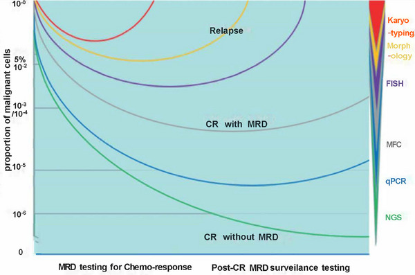
Minimal residual disease (MRD) serves as a pivotal biomarker for the clinical diagnosis and subsequent treatment of cancer patients. In hematological malignancies, MRD pose an increasingly serious threat to the health of Chinese people. Accurate MRD detection is essential for assessing relapse risk and optimizing therapeutic strategies, yet current methods such as flow cytometry, polymerase chain reaction (PCR), and next-generation sequencing (NGS) each have distinct limitations, and significant gaps remain in achieving optimal sensitivity and specificity of these technologies. This review provides a comprehensive analysis of MRD detection methods, high-lighting their clinical implications, including their roles in treatment decision-making, risk stratification, and patient outcomes. It discusses the strengths and weaknesses of existing techniques and explores emerging technologies that promise enhanced diagnostic precision. Key advancements such as integrating NGS with other methodologies and novel approaches like liquid biopsy and PCR are examined. The review underscores the academic and practical value of early and accurate MRD detection, emphasizing its impact on improving patient management and treatment outcomes. By addressing the limitations of current technologies and exploring future directions, this review aims to advance the field and support personalized medicine approaches to cancer treatment.

Keywords: MRD; clinical implication; detection methods; next‐generation sequencing.


Conflict of interest statement

The authors declare no conflicts of interest.

Figures

FIGURE 1
FIGURE 1
Results of treatment response testing and relapse patterns in patients with minimal residual disease (MRD) using different techniques with varying sensitivity levels. MRD detection is critical for monitoring treatment response and predicting relapse risk. A deeper response correlates with a better prognosis. However, even patients who achieve complete remission (CR) may relapse, suggesting that residual cancer cells may still be present. The level of MRD is closely associated with relapse risk. Abbreviations: complete remission (CR); fluorescence in situ hybridization (FISH); multiparameter flow cytometry (MFC); quantitative polymerases chain reaction (qPCR); next‐generation sequencing (NGS); minimal residual disease (MRD).
FIGURE 2
FIGURE 2
Technical principle of fluorescence in situ hybridization (FISH). FISH uses fluorescently labeled DNA probes to hybridize with specific DNA or RNA sequences in the sample, allowing for the detection and localization of genetic material within cells.
FIGURE 3
FIGURE 3
Workflow of next‐generation sequencing (NGS) process. NGS involves adding adapters to fragmented nucleic acids, amplifying them with complementary primers, enriching the library, performing sequencing, and analyzing the results through signal scanning.
FIGURE 4
FIGURE 4
Evaluation of NGS‐based MRD detection in CD34+/CD117+ cells. This figure compares NGS‐based MRD detection with donor chimerism (DC) analysis, highlighting sensitivity in peripheral blood (PB) and bone marrow (BM) samples. NGS‐based detection shows high correlation with DC analysis in sorted CD34+/CD117+ PB cells, with better sensitivity when initial FACS enrichment was performed. (A) Correlation of MRD detection using NGS or DC analysis in sorted CD34+/CD117+ PB cell samples. (B) NGS‐based MRD positivity rates in relation to the corresponding CD34+/CD117+ DC level in PB. The cutoff for NGS‐based MRD quantification is indicated at 0.01% VAF. (C) Detection of the Kasumi cell line in PB using NGS‐based quantification of the KIT N822K variant (red dots) or by CD34+ DC analysis (blue dots). (D) Correlation of NGS‐based MRD detection in sorted CD34+ cells of matched PB and BM samples as templates for analysis. (E) Quantification of variant allele frequencies (%) in CD34+ cells using matched PB or BM samples for NGS. (F) Quantification of mutant alleles by NGS using sorted CD34+/CD117+ PB cells or unsorted material of matched follow‐up samples. Box plots represent median values with interquartile range; box whiskers represent minimum to maximum values. Abbreviations: next‐generation sequencing (NGS); minimal residual disease (MRD); donor chimerism (DC); peripheral blood (PB); variant allele frequency (VAF). Reproduced with permission from Ref. [79], Copyright © 2022 The American Society of Hematology.
FIGURE 5
FIGURE 5
Kaplan–Meier analysis of PFS and OS of MRD‐positive versus MRD‐negative. Patients Using NGS and NGF. Kaplan–Meier curves compare progression free survival (PFS) and overall survival (OS) between MRD‐positive and MRD‐negative subsets, revealing no significant differences in detection results between NGS and NGF. The analysis demonstrates that MRD‐negative patients have significantly better PFS and OS rates than MRD‐positive patients, when time calculated from MRD assessment 3 months posttransplantation. Negative patients are indicated in black, while positive patients are shown in red, with patient risk levels detailed at each time point. Events are represented between parentheses. (A) PFS of NGS‐based results. (B) PFS of NGF‐based results. (C) OS of NGS‐based results. (D) OS of NGF‐based results. Abbreviations: next‐generation sequencing (NGS); next‐generation flow (NGF); minimal residual disease (MRD). Reproduced with permission from Ref. [144], Copyright © 2020, The Author(s).

Similar articles

References

    1. Liu W., Liu J., Song Y., et al., “Mortality of Lymphoma and Myeloma in China, 2004–2017: An Observational Study,” Journal of Hematology & Oncology 12, no. 1 (2019): 22. - PMC - PubMed
    1. Chinese Society of Immunology, Clinical Flow Cytometry Group . Expert Consensus on Minimal Residual Disease Detection of Acute Leukemia and Plasma Cell Neoplasms by Multi‐parameter Flow Cytometry. Zhonghua Xue Ye Xue Za Zhi = Zhonghua Xueyexue Zazhi 2017;38(12):1001–1011. - PMC - PubMed
    1. Bernardi M., Ferrara F., Carrabba M. G., et al., “MRD in Venetoclax‐Based Treatment for AML: Does It Really Matter?,” Frontiers in Oncology 12 (2022): 890871. - PMC - PubMed
    1. Yang F., Anekpuritanang T., and Press R. D., “Clinical Utility of Next‐Generation Sequencing in Acute Myeloid Leukemia,” Molecular Diagnosis & Therapy 24, no. 1 (2020): 1–13. - PubMed
    1. Li W.. Measurable Residual Disease Testing in Acute Leukemia: Technology and Clinical Significance. In: Li W, ed. “Leukemia” (Brisbane (AU): Exon Publications, 2022). October 16,. - PubMed

LinkOut - more resources