

Effects of stellate ganglion block on inflammation and autophagy of spinal cord neurons in rats with neuropathic pain after spinal cord injury
- PMID: 40385057
- PMCID: PMC12082548
- DOI: 10.62347/QEVD3665
Effects of stellate ganglion block on inflammation and autophagy of spinal cord neurons in rats with neuropathic pain after spinal cord injury
Abstract
Objective: To assess the therapeutic effects of stellate ganglion block (SGB) on spinal cord injury (SCI)-induced neuropathic pain in rats, and to explore its potential mechanisms in alleviating neuropathic pain, thereby providing a theoretical foundation for clinical treatment.
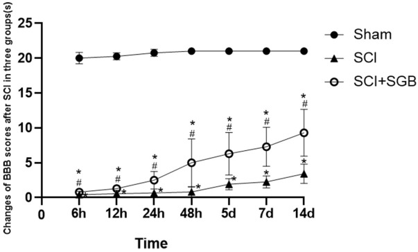
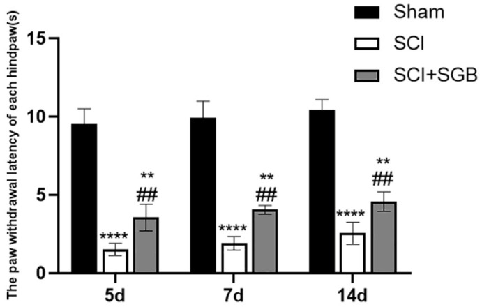
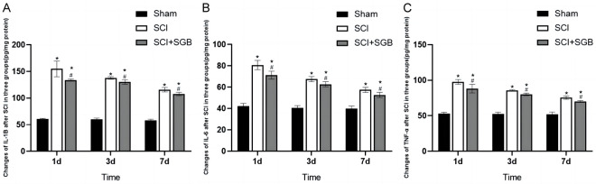
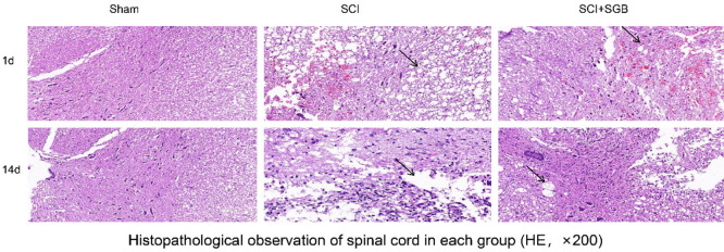
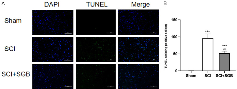
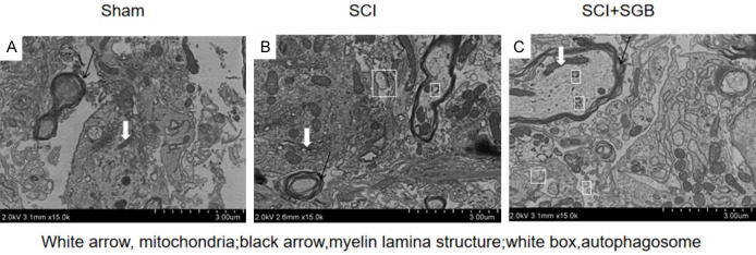
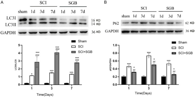
Methods: A rat model of SCI was established, and animals were randomly assigned to one of three groups: the sham surgery group (Sham), the SCI group (SCI), or the SCI group treated with SGB (SCI + SGB). Motor function was assessed using the Basso Beattie Bresnahan (BBB) locomotor rating scale, while thermal hyperalgesia was evaluated using hot plate test. Enzyme-linked immunosorbent assay (ELISA) was utilized to measure the levels of inflammatory cytokines, including interleukin-1β (IL-1β), IL-6, and tumor necrosis factor-α (TNF-α), within the spinal cord. Hematoxylin-eosin (HE) staining was performed to observe spinal cord histopathology. Terminal deoxynucleotidyl transferase-mediated dUTP nick-end labeling (TUNEL) staining was used to detect apoptotic cells, and transmission electron microscopy was employed to visualize autophagosomes. Expression of autophagy-related proteins LC3-II/LC3-I and p62 was examined via Western blotting.
Results: Compared with the sham group, rats in the SCI group displayed impaired hind limb motor function, decreased pain thresholds, elevated inflammatory cytokine levels, significant spinal cord pathology, increased apoptosis, altered expression of autophagy-related protein, and disrupted autophagic flux. In contrast, SGB treatment improved motor function, alleviated pain, reduced inflammatory cytokines levels, mitigated spinal cord injury and apoptosis, and enhanced autophagy with improved autophagic flux.
Conclusions: Stellate ganglion block alleviates neuropathic pain in SCI-induced rats by reducing pro-inflammatory cytokine levels, mitigating spinal cord apoptosis and injury, promoting autophagy, and restoring autophagic flux in the spinal cord.
Keywords: Stellate ganglion block; neuropathic pain; spinal cord injury.
AJTR Copyright © 2025.
Conflict of interest statement
None.
Figures
Similar articles
-
[Expression of B-cell lymphoma-2 protein multisite phosphorylation in autophagy after spinal cord injury in rats].Zhongguo Xiu Fu Chong Jian Wai Ke Za Zhi. 2019 May 15;33(5):618-627. doi: 10.7507/1002-1892.201812064. Zhongguo Xiu Fu Chong Jian Wai Ke Za Zhi. 2019. PMID: 31090358 Free PMC article. Chinese.
-
Cinepazide maleate promotes recovery from spinal cord injury by inhibiting inflammation and prolonging neuronal survival.Drug Dev Res. 2023 Jun;84(4):736-746. doi: 10.1002/ddr.22052. Epub 2023 Mar 29. Drug Dev Res. 2023. PMID: 36988113
-
Overexpression of HIPK2 attenuates spinal cord injury in rats by modulating apoptosis, oxidative stress, and inflammation.Biomed Pharmacother. 2018 Jul;103:127-134. doi: 10.1016/j.biopha.2018.03.117. Epub 2018 Apr 24. Biomed Pharmacother. 2018. PMID: 29649627
-
Local Delivery of β-Elemene Improves Locomotor Functional Recovery by Alleviating Endoplasmic Reticulum Stress and Reducing Neuronal Apoptosis in Rats with Spinal Cord Injury.Cell Physiol Biochem. 2018;49(2):595-609. doi: 10.1159/000492996. Epub 2018 Aug 30. Cell Physiol Biochem. 2018. PMID: 30165357
-
A study on the mechanism of PP2A in the recovery of SCI in rats through downregulation of MMP-9 via MAPK signaling pathway.Eur Rev Med Pharmacol Sci. 2021 Dec;25(23):7195-7203. doi: 10.26355/eurrev_202112_27411. Eur Rev Med Pharmacol Sci. 2021. PMID: 34919217
References
-
- Widerström-Noga E. Neuropathic pain and spinal cord injury: management, phenotypes, and biomarkers. Drugs. 2023;83:1001–1025. - PubMed
LinkOut - more resources
Full Text Sources