. 2025 May 18;18(5):792-801.
doi: 10.18240/ijo.2025.05.04. eCollection 2025.

Fenofibrate mitigates the dysfunction of high glucose-driven human retinal microvascular endothelial cells by suppressing NLRP3 inflammasome

Affiliations

Fenofibrate mitigates the dysfunction of high glucose-driven human retinal microvascular endothelial cells by suppressing NLRP3 inflammasome

Yi Shi et al. Int J Ophthalmol. .

Abstract

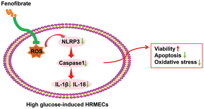
Aim: To determine the therapeutic benefits of fenofibrate (Feno) on the dysfunction of high glucose (HG)-induced human retinal microvascular endothelial cells (HRMECs) and to elucidate the underlying molecular mechanism.

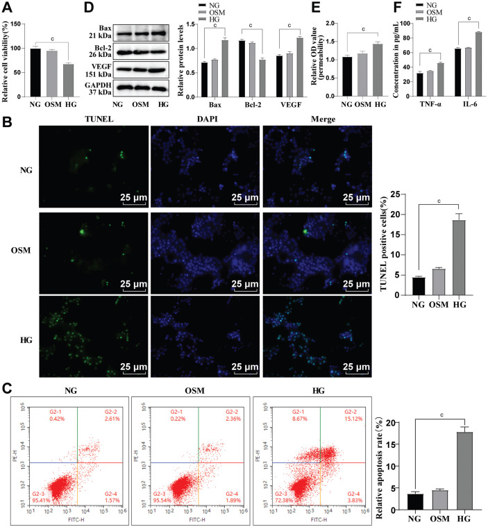
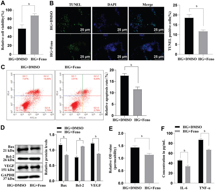
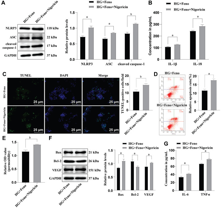
Methods: HRMEC dysfunction model was established by 48h glucose (30 mmol/L) treatment and treated with Feno/NOD-like receptor thermal protein domain associated protein 3 (NLRP3) inflammasome activator (Nigericin). Cell viability/apoptosis were assessed by cell counting kit-8 (CCK-8)/terminal deoxynucleotidyl transferase-mediated dUTP-biotin nick end labeling assay (TUNEL) staining and flow cytometry assays. Levels of apoptosis- (Bcl-2-associated X protein, Bax/B-cell lymphoma 2, Bcl-2), vascular permeability-(vascular endothelial growth factor, VEGF) and inflammasome activation-related proteins (NLRP3/cleaved caspase-1/apoptosis-associated speck-like protein containing a CARD, ASC), as well as inflammatory factors (interleukin, IL-6/IL-1β/tumor necrosis factor, TNF-α/IL-18) were determined with Western blot/enzyme linked immunosorbent assay (ELISA). Cell permeability/reactive oxygen species (ROS) level/superoxide dismutase (SOD) activity/malondialdehyde (MDA) content were assessed by Evans blue staining/2',7'-dichlorodihydrofluorescein diacetate (DCFH-DA) fluorescent probe/SOD kit/MDA kit.

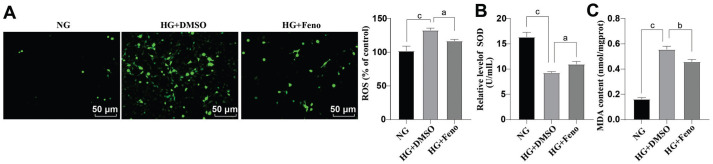
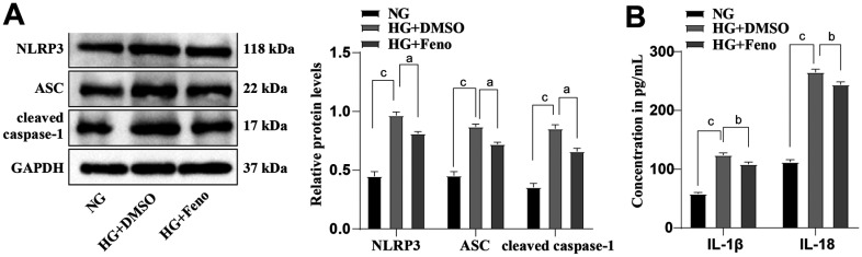
Results: HRMEC dysfunction was successfully induced by HG, evidenced by decreased viability (P<0.001), increased apoptosis (P<0.001), permeability (P<0.001), and inflammatory factor levels (P<0.001). Feno treatment significantly ameliorated HG-induced HRMEC dysfunction (P<0.01). Meanwhile, HG induction increased ROS production (P<0.001) and MDA content (P<0.001) in HRMECs, while reducing SOD activity (P<0.001), indicative of oxidative stress. This was, however, abolished by Feno (P<0.05). Moreover, Feno eliminated activation of NLRP3 inflammasomes (P<0.05) in HG-induced HRMECs. Strikingly, activation of NLRP3 inflammasomes partially averted the inhibition of Feno on HG-induced HRMEC dysfunction (P<0.05).

Conclusion: Feno represses oxidative stress and NLRP3 inflammasome activation, consequently alleviating HG-induced HRMEC dysfunction.

Keywords: NOD-like receptor thermal protein domain associated protein 3 inflammasomes; fenofibrate; high glucose; human retinal microvascular endothelial cells; oxidative stress.


Conflict of interest statement

Conflicts of Interest: Shi Y, None; Chen HM, None; Liu AH, None; Li XR, None.

Figures

Figure 1
Figure 1. The influence of HG on apoptosis of HRMECs
A: CCK-8 was conducted to evaluate cell viability; B: TUNEL staining was conducted to assess apoptosis; C: Flow cytometry was adopted to evaluate apoptosis; D: Western blot was conducted to measure the protein levels of apoptosis-associated proteins Bax and Bcl-2, as well as vascular permeability-related protein VEGF; E: Cell permeability was assayed by Evans blue staining; F: ELISA was conducted to measure TNF-α and IL-6 levels. The cell-based in vitro experiments were run three times in an independent manner, with the data represented as mean±SD. Multi-group data were compared by one-way ANOVA, with Tukey's test. cP<0.001. HRMECs: Human retinal microvascular endothelial cells; NG: Normal glucose; HG: High glucose; OSM: Osmotic pressure; CCK-8: Cell counting kit-8; DMSO: Dimethyl sulphoxide; TUNEL: Terminal deoxynucleotidyl transferase-mediated dUTP-biotin nick end labeling; TNF-α: Tumor necrosis factor-α; IL-6: Interleukin-6; ELISA: Enzyme-linked immunosorbent assay; GAPDH: Glyceraldehyde-3-phosphate dehydrogenase; Bax: Bcl-2-associated X protein; Bcl-2: B-cell lymphoma 2; VEGF: Vascular endothelial growth factor; OD: Optical density; DAPI: 4′,6-diamidino-2-phenylindole; SD: Standard deviation; ANOVA: Analysis of variance.
Figure 2
Figure 2. The role of Feno in HG-induced dysfunction of HRMECs
A: CCK-8 was performed to determine cell viability; B: TUNEL staining was performed to evaluate cell apoptosis; C: The cell apoptosis was assessed by flow cytometry; D: Determination of Bax, Bcl-2, and VEGF protein levels by Western blot; E: Evaluation of cell permeability by Evans blue staining; F: ELISA assay was performed to assess the levels of IL-6 and TNF-α. The cell-based in vitro experiments were run three times in an independent manner. The data were represented as mean±SD, with independent sample t-test adopted for two-group data comparisons. bP<0.01. CCK-8: Cell counting kit-8; HG: High glucose; Feno: Fenofibrate; HRMECs: Human retinal microvascular endothelial cells; DAPI: 4′,6-diamidino-2-phenylindole; DMSO: Dimethyl sulphoxide; TUNEL: Terminal deoxynucleotidyl transferase-mediated dUTP-biotin nick end labeling; Bax: Bcl-2-associated X protein; TNF-α: Tumor necrosis factor-α; IL-6: Interleukin-6; OD: Optical density; GAPDH: Glyceraldehyde-3-phosphate dehydrogenase; VEGF: Vascular endothelial growth factor; Bcl-2: B-cell lymphoma 2; SD: Standard deviation; ELISA: Enzyme-linked immunosorbent assay.
Figure 3
Figure 3. Influence of Feno on HG-induced oxidative stress
A: Fluorescent probe DCFH-DA was adopted to detect intracellular ROS; B: SOD activity measurement was done by the SOD kit; C: The MDA content measurement was done by an MDA kit. The cell-based in vitro experiments were run three times in an independent manner, and the data were represented as mean±SD. Data were analyzed by virtue of one-way ANOVA with Tukey's test. aP<0.05, bP<0.01, cP<0.001. Feno: Fenofibrate; SOD: Superoxide dismutase; NG: Normal glucose; DMSO: Dimethyl sulphoxide; ROS: Reactive oxygen species; MDA: Malondialdehyde; HG: High glucose; ANOVA: Analysis of variance; DCFH-DA: 2′,7′-Dichlorodihydrofluorescein diacetate.
Figure 4
Figure 4. Influence of Feno on the activation of NLRP3 inflammasomes
A: Western blot was performed to measure the protein levels of ASC, NLRP3 and cleaved caspase-1; B: ELISA was performed to analyze the levels of IL-18 and IL-1β. The cell-based in vitro experiments were run three times in an independent manner, and the data were represented as the mean±SD. Data were analyzed by one-way ANOVA with Tukey's test. aP<0.05, bP<0.01, cP<0.001. Feno: Fenofibrate; NLRP3: NOD-like receptor thermal protein domain-associated protein 3; ASC: Apoptosis-associated speck-like protein containing a CARD; GAPDH: Glyceraldehyde-3-phosphate dehydrogenase; HG: High glucose; IL: Interleukin; SD: Standard deviation; DMSO: Dimethyl sulphoxide; ELISA: Enzyme-linked immunosorbent assay; NG: Normal glucose; ANOVA: Analysis of variance.
Figure 5
Figure 5. The effect of activating NLRP3 inflammasomes on the prevention of Feno on HRMEC dysfunction
A: Detection of ASC, NLRP3, and cleaved caspase-1 protein levels were done by virtue of Western blot; B: ELISA was conducted to analyze IL-1β and IL-18 levels; C: TUNEL assay was conducted to assess HRMEC apoptosis; D: Flow cytometry was conducted to evaluate HRMEC apoptosis; E: Cell permeability was tested by Evans blue staining; F: Determination of Bax, Bcl-2 and VEGF protein levels by Western blot; G: ELISA was conducted to measure the levels of IL-6 and TNF-α. The cell-based in vitro experiments were run three times in an independent manner, with the data represented as mean±SD. Independent sample t-test was performed to analyze. aP<0.05, bP<0.01. Feno: Fenofibrate; DAPI: 4′,6-diamidino-2-phenylindole; NLRP3: NOD-like receptor thermal protein domain-associated protein 3; HRMEC: Human retinal microvascular endothelial cell; HG: High glucose; ASC: Apoptosis-associated speck-like protein containing a CARD; DMSO: Dimethyl sulphoxide; TNF-α: Tumor necrosis factor-α; IL: Interleukin; ELISA: Enzyme-linked immunosorbent assay; Bcl-2: B-cell lymphoma 2; VEGF: Vascular endothelial growth factor; OD: Optical density; Bax: Bcl-2-associated X protein; SD: Standard deviation.
Figure 6
Figure 6. Schematic representation summarizing the role of Feno in HG-induced dysfunction of HRMECs
Feno reduces ROS generation, increases SOD activity, and suppresses NLRP3 inflammasome activation, which in turn decreases caspase-1 activation and pro-inflammatory cytokine release. This process plays a key role in reducing inflammation, oxidative stress, and cellular damage under HG conditions. SOD: Superoxide dismutase; NLRP3: NOD-like receptor protein 3; Caspase-1: Cysteine-dependent aspartate-specific protease 1; ROS: Reactive oxygen species; IL-18: Interleukin-18; IL-1β: Interleukin-1β; HRMEC: Human retinal microvascular endothelial cells.

Similar articles

References

    1. Roy S, Kim D. Retinal capillary basement membrane thickening: Role in the pathogenesis of diabetic retinopathy. Prog Retin Eye Res. 2021;82:100903. - PMC - PubMed
    1. Antonetti DA, Silva PS, Stitt AW. Current understanding of the molecular and cellular pathology of diabetic retinopathy. Nat Rev Endocrinol. 2021;17(4):195–206. - PMC - PubMed
    1. Fang XL, Zhang Q, Xue WW, et al. Suppression of cAMP/PKA/CREB signaling ameliorates retinal injury in diabetic retinopathy. Kaohsiung J Med Sci. 2023;39(9):916–926. - PMC - PubMed
    1. Li Y, Li HY, Shao J, et al. GRP75 modulates endoplasmic reticulum–mitochondria coupling and accelerates Ca2+-dependent endothelial cell apoptosis in diabetic retinopathy. Biomolecules. 2022;12(12):1778. - PMC - PubMed
    1. Magupalli VG, Negro R, Tian YZ, et al. HDAC6 mediates an aggresome-like mechanism for NLRP3 and pyrin inflammasome activation. Science. 2020;369(6510):eaas8995. - PMC - PubMed

LinkOut - more resources