

Femtosecond Laser Direct Writing of Diffraction Gratings for Modifying the Refractive Index of Intraocular Lenses
- PMID: 40385207
- PMCID: PMC12079213
- DOI: 10.1021/acsomega.4c11380
Femtosecond Laser Direct Writing of Diffraction Gratings for Modifying the Refractive Index of Intraocular Lenses
Abstract
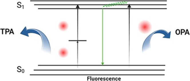
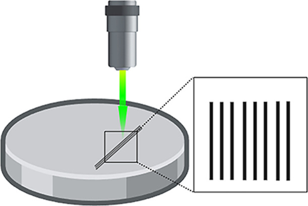
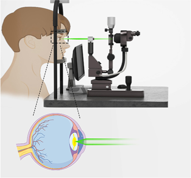
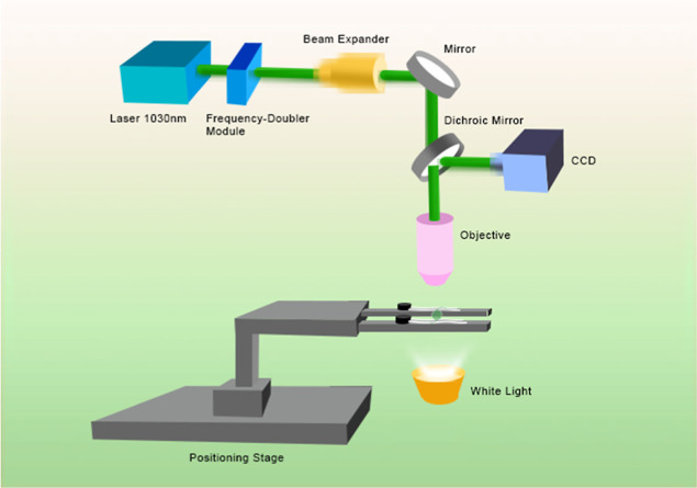
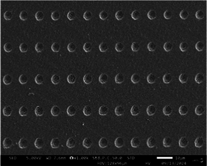
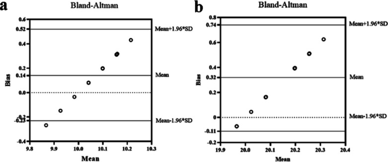
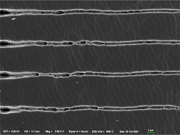
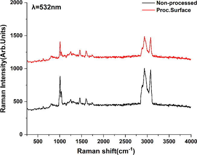
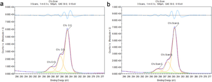
An advanced femtosecond laser experimental platform with high precision was developed for the reconstruction of the refractive index of intraocular lenses (IOLs), and its accuracy was rigorously evaluated. Diffraction gratings were inscribed on the surface of an acrylate sample utilizing a fiber femtosecond laser operating at a wavelength of 515 nm with a repetition rate of 40 MHz. The samples were subsequently measured using an Abbe refractometer to assess the alterations in their refractive index induced by the femtosecond laser scanning process. Scanning electron microscopy, confocal Raman microscopy, and X-ray photoelectron spectroscopy were employed to examine the morphology of the diffraction grating on the sample surface following femtosecond laser scanning. Additionally, these techniques were utilized to investigate the alterations in molecular structure within the material postlaser scanning, as well as to elucidate the underlying mechanisms responsible for changes in refractive index. Furthermore, the parameters of the femtosecond laser utilized in this study were compared with those of lasers commonly employed in clinical settings.
© 2025 The Authors. Published by American Chemical Society.
Conflict of interest statement
The authors declare no competing financial interest.
Figures
Similar articles
-
Femtosecond laser direct writing of diffraction grating and its refractive index change in chalcogenide As2Se3 film.Opt Express. 2019 Oct 14;27(21):30090-30101. doi: 10.1364/OE.27.030090. Opt Express. 2019. PMID: 31684262
-
The Role of Thermal Accumulation on the Fabrication of Diffraction Gratings in Ophthalmic PHEMA by Ultrashort Laser Direct Writing.Polymers (Basel). 2020 Dec 11;12(12):2965. doi: 10.3390/polym12122965. Polymers (Basel). 2020. PMID: 33322569 Free PMC article.
-
High-Repetition-Rate Femtosecond Laser Processing of Acrylic Intra-Ocular Lenses.Polymers (Basel). 2020 Jan 20;12(1):242. doi: 10.3390/polym12010242. Polymers (Basel). 2020. PMID: 31968562 Free PMC article.
-
Intratissue refractive index shaping (IRIS) of the cornea and lens using a low-pulse-energy femtosecond laser oscillator.Invest Ophthalmol Vis Sci. 2008 Dec;49(12):5332-9. doi: 10.1167/iovs.08-1921. Epub 2008 Jul 18. Invest Ophthalmol Vis Sci. 2008. PMID: 18641284 Free PMC article.
-
Ultrafast Laser Processing of Optical Fibers for Sensing Applications.Sensors (Basel). 2021 Feb 19;21(4):1447. doi: 10.3390/s21041447. Sensors (Basel). 2021. PMID: 33669717 Free PMC article. Review.
References
LinkOut - more resources
Full Text Sources