. 2025 May 2:13:1598736.
doi: 10.3389/fcell.2025.1598736. eCollection 2025.

Combined use of olfactory mucosal mesenchymal stem cells conditioned medium and neural guide conduits promotes nerve regeneration in an ovine model

Affiliations

Combined use of olfactory mucosal mesenchymal stem cells conditioned medium and neural guide conduits promotes nerve regeneration in an ovine model

Rui Alvites et al. Front Cell Dev Biol. .

Abstract

Introduction: Peripheral nerve injuries remain a significant clinical challenge, particularly in severe neurotmesis injuries requiring complex therapeutic interventions to restore functionality. This study aimed to evaluate the pro-regenerative potential of combining neural guide conduits with conditioned medium from olfactory mucosa mesenchymal stem cells, compared to gold-standard surgical techniques.

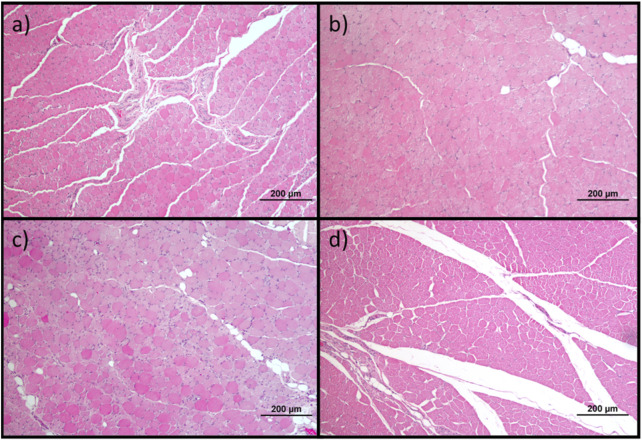
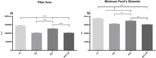
Methods: The study was conducted using a validated ovine model of common peroneal nerve injury. Recovery was assessed over 24 weeks through functional, kinematic, ultrasonographic, and electrophysiological evaluations, complemented by post-mortem nerve stereology and muscle histomorphometry.

Results: All therapeutic approaches promoted nerve and muscle regeneration, resulting in notable functional and structural improvements. However, irregularities were observed, as neural guide conduits and conditioned medium did not consistently outperform standard techniques. Additionally, recovery often fell short of normal values in the control group.

Discussion: These findings highlight the complexity of peripheral nerve regeneration in challenging surgical scenarios and underscore the translational potential of biomaterials and cell conditioned medium-based therapies. However, the observed irregularities emphasize the need for further research in complex animal models before application in real clinical cases. Such studies are essential to refine therapeutic strategies, address inconsistencies, and establish cell conditioned medium as a viable tool in peripheral nerve regeneration and repair.

Keywords: animal model; cell-based therapies; common peroneal nerve; conditioned medium; olfactory mucosa mesenchymal stem cells; peripheral nerve injury; peripheral nerve regeneration; sheep model.


Conflict of interest statement

The authors declare that the research was conducted in the absence of any commercial or financial relationships that could be construed as a potential conflict of interest. The author(s) declared that they were an editorial board member of Frontiers, at the time of submission. This had no impact on the peer review process and the final decision.

Figures

FIGURE 1
FIGURE 1
Experimental therapies applied to the common peroneal nerve after neurotmesis injury. The applied NGCs were 30 mm long and had an internal diameter of 3 mm.
FIGURE 2
FIGURE 2
Results of posture evaluation performed in stationary position on all animals over the study period of 24 weeks. UC: Uninjured Control; EtE: end-to-end suture; NGC: application of Reaxon® NGC; NGC-CM: application of Reaxon® NGC and OM-MSCs CM. Classification key - 1: Digits and hock in physiological position, no postural changes; 2: Mild flexion of digits and/or extension of the hock; 3: Moderated flexion of digits and/or extension of the hock; 4: Pronounced flexion of digits and extension of the hock; 5: Severe flexion of digits and extension of the hock. Results presented as Mean +SEM.
FIGURE 3
FIGURE 3
Stationary posture of animals from the NGC group at two different timepoints: (a) posture at T1: overflexion of the distal joints and overextension of the hock, with the dorsal surfaces of the digits in contact with the ground (red arrow); (b) posture at T24: digits and hock in physiological position, with the plantar surface of the digits in contact with the ground and without postural changes (blue arrow).
FIGURE 4
FIGURE 4
Results of postural reactions evaluation using: (a) proprioceptive assessment through static repositioning over the study period of 24 weeks; (b) proprioceptive assessment through dynamic repositioning over the study period of 24 weeks. UC: Uninjured Control; EtE: end-to-end suture; NGC: application of Reaxon® NGC; NGC-CM: application of Reaxon® NGC and OM-MSCs CM. Classification key—1: <3 s; 2: 3–5 s; 3: 5–10 s; 4: 10–15 s; 5: 15–20 s; 6: >20 s. Results presented as Mean +SEM.
FIGURE 5
FIGURE 5
Results of spinal reflexes assessed using the withdrawal reflex over the study period of 24 weeks. UC: Uninjured Control; EtE: end-to-end suture; NGC: application of Reaxon® NGC; NGC-CM: application of Reaxon® NGC and OM-MSCs CM. Classification key - Spinal Reflexes - 1: Absent reflex; 2: Reflex present but delayed; 3: Reflex present. Results presented as Mean +SEM.
FIGURE 6
FIGURE 6
Results of kinematic evaluation performed at 24 weeks. Upper graphs show the ankle (A1), knee (A2) and hip (A3) angles during the stance and swing phases of the gait cycle. The lower graphs show SPM statistic as a function of the gait cycle: (B1), (B2), (B3) - ETS vs. UC for ankle, knee and hip, respectively; (C1), (C2), (C3) – NGC vs. UC for ankle, knee and hip, respectively; and (D1), (D2), (D3) – NGC-CM vs. UC for ankle, knee and hip, respectively. The moments of the gait cycle in which the critical threshold (t ∗) was exceeded are represented by the grey area of the lower graphs.
FIGURE 7
FIGURE 7
Ultrasonographic appearance of the common peroneal nerves submitted to different treatments. (a) intact common peroneal nerve (UC); (b–d) nerves that received an EtE suture 1 month, 3 months and 6 months after injury, respectively; (e–g) nerves that received a NGC 1 month, 3 months and 6 months respectively; (h) nerve that received a NGC 6 months after surgery, transversal section; (i–k) nerves that received the combination NGC-CM 1 month, 3 months and 6 months after injury, respectively; (l) nerve that received the combination NGC-CM 6 months after surgery, transversal section. In each panel, the red arrow indicates the common peroneal nerve or common peroneal nerve + NGC set.
FIGURE 8
FIGURE 8
Results of ultrasound measurements of the diameter of the intervened common peroneal nerves over the 24-weeks study period. UC: Uninjured Control; EtE: end-to-end suture; NGC: application of Reaxon® NGC; NGC-CM: application of Reaxon® NGC and OM-MSCs CM. Results presented as Mean ± SEM.
FIGURE 9
FIGURE 9
Ultrasonographic appearance of the cranial tibial muscle as an effector muscle of the injured common peroneal nerves subject to different treatments: (a) intact common peroneal nerve (UC); (b–d) nerves that received an EtE suture 1 month, 3 months and 6 months after injury, respectively; (e–g) nerves that received a NGC 1 month, 3 months and 6 months respectively; (h–j) nerves that received the combination NGC-CM 1 month, 3 months and 6 months after injury, respectively. The red dashed circle delimits the muscle, in which the thickness (vertical red dashed line) and width (horizontal red dashed line) were measured. 1- Cranial tibial muscle; 2- long digital extensor muscle; 3- tibia.
FIGURE 10
FIGURE 10
Results of ultrasound measurements of the width and thickness of the cranial tibial muscle over the 24-weeks study period. UC: Uninjured Control; EtE: end-to-end suture; NGC: application of Reaxon® NGC; NGC-CM: application of Reaxon® NGC and OM-MSCs CM. Results presented as Mean +SEM.
FIGURE 11
FIGURE 11
Results of amplitude and latency determined in the cranial tibial muscle over the 24-week study period, through electrophysiological assessment. UC: Uninjured Control; EtE: end-to-end suture; NGC: application of Reaxon® NGC; NGC-CM: application of Reaxon® NGC and OM-MSCs CM. Results presented as Mean +SEM.
FIGURE 12
FIGURE 12
Results of the stereological assessment of the common peroneal nerve 24 weeks after neurotmesis: (a) density of fibers; (b) total number of fibers; (c) axon diameter; (d) fiber diameter; (e) myelin thickness; (f) g-ratio (mean ± SEM)). * corresponds to 0.01 ≤ p < 0.05, ** to 0.001 ≤ p < 0.01, *** to 0.0001 ≤ p < 0.001, and **** to p < 0.0001.
FIGURE 13
FIGURE 13
Toluidine blue-stained images of common peroneal nerve semi-thin sections of the different therapeutic groups 24 weeks after surgery: (A) UC; (B) EtE; (C) NGC; (D) NGC-CM; Scale bars = 20 µm.
FIGURE 14
FIGURE 14
Histological images of the cranial tibial muscles subjected to histomorphometric analysis in the different groups: (a) UC; (b) EtE; (c) NGC; (d) NGC-CM; Magnifications: ×100.
FIGURE 15
FIGURE 15
Histomorphometric analysis of cranial tibial muscle: (a) individual fiber area; (b) minimum Feret’s diameter of the muscle fibers (mean ± SEM). ** to 0.001 ≤ p < 0.01, and **** to p < 0.0001.
FIGURE 16
FIGURE 16
Percentage of muscle mass lost in each therapeutic group as a function of contralateral healthy muscle weight (mean ± SEM).

Similar articles

References

    1. Aisaiti A., Aierxiding S., Shoukeer K., Muheremu A. (2024). Mesenchymal stem cells for peripheral nerve injury and regeneration: a bibliometric and visualization study. Front. Neurol. 15, 1420402. 10.3389/fneur.2024.1420402 - DOI - PMC - PubMed
    1. Alvites R., Branquinho M., Sousa A. C., Lopes B., Sousa P., Mauricio A. C. (2022a). Mesenchymal stem/stromal cells and their paracrine activity-immunomodulation mechanisms and how to influence the therapeutic potential. Pharmaceutics 14 (2), 381. 10.3390/pharmaceutics14020381 - DOI - PMC - PubMed
    1. Alvites R., Lopes B., Coelho A., Maurício A. C. (2024). Peripheral nerve regeneration: A challenge far from being overcome. Regenerative Medicine 19 (4), 155–159. 10.2217/rme-2023-0072 - DOI - PubMed
    1. Alvites R., Lopes B., Sousa P., Sousa A. C., Coelho A., Moreira A., et al. (2023). Ultrasound landmarks in the approach to the common peroneal nerve in a sheep model-application in peripheral nerve regeneration. Life (Basel) 13 (9), 1919. 10.3390/life13091919 - DOI - PMC - PubMed
    1. Alvites R., Rita Caseiro A., Santos Pedrosa S., Vieira Branquinho M., Ronchi G., Geuna S., et al. (2018a). Peripheral nerve injury and axonotmesis: state of the art and recent advances. Cogent Med. 5 (1), 1466404. 10.1080/2331205x.2018.1466404 - DOI

LinkOut - more resources