. 2025 Apr 25;5(1):ycaf070.
doi: 10.1093/ismeco/ycaf070. eCollection 2025 Jan.

Photosynthetic dependence and filament production in physical bacterial-Symbiodiniaceae interactions

Affiliations

Photosynthetic dependence and filament production in physical bacterial-Symbiodiniaceae interactions

Gavin C McLaren et al. ISME Commun. .

Abstract

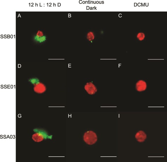
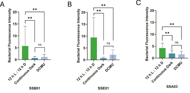
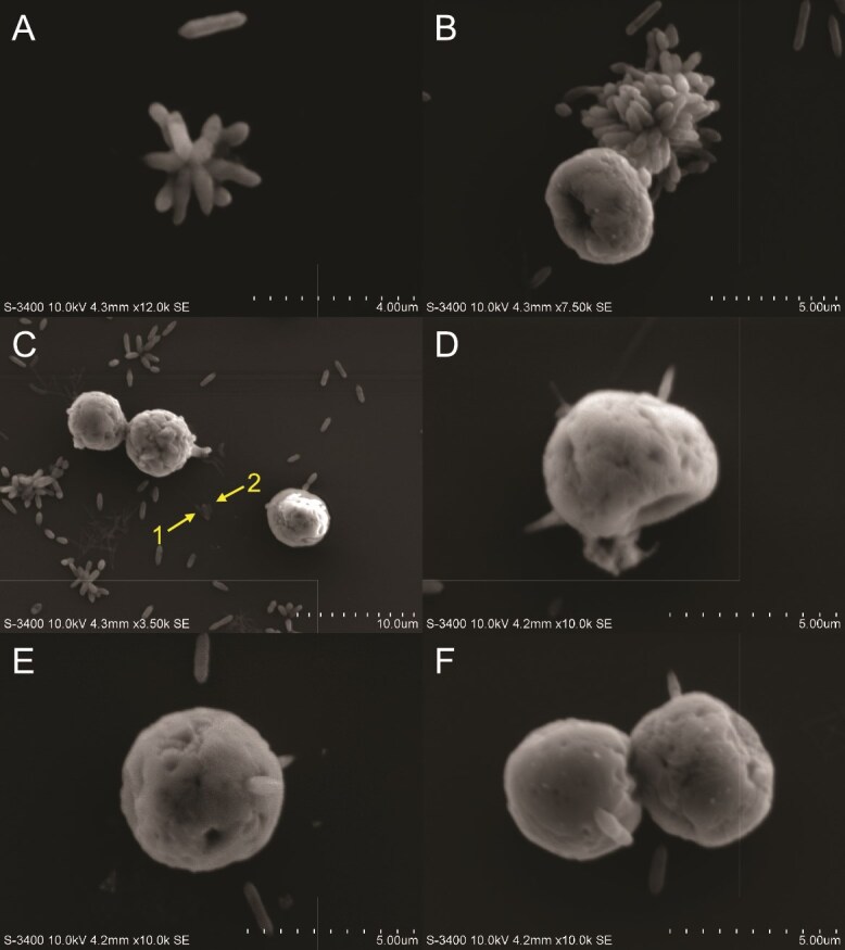
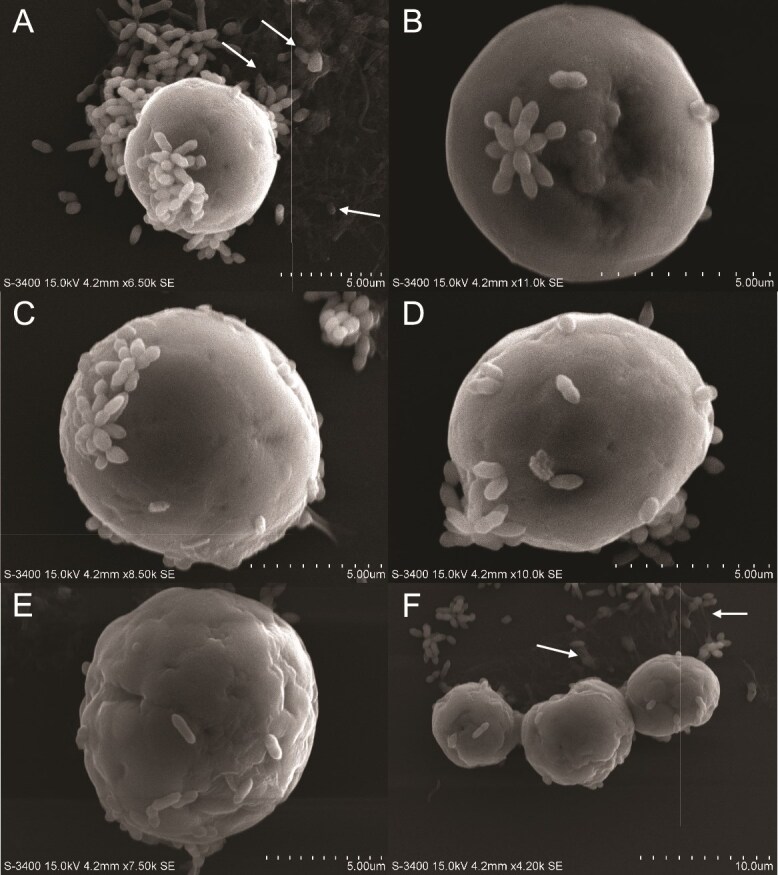
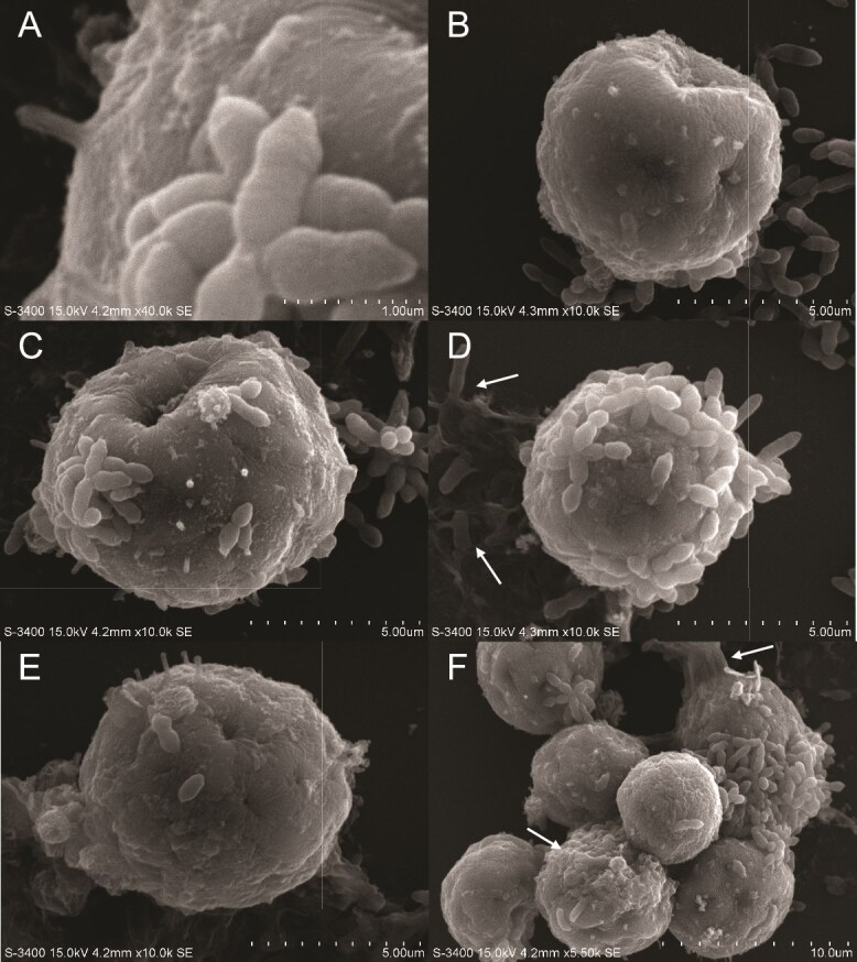
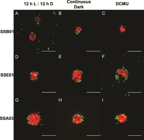
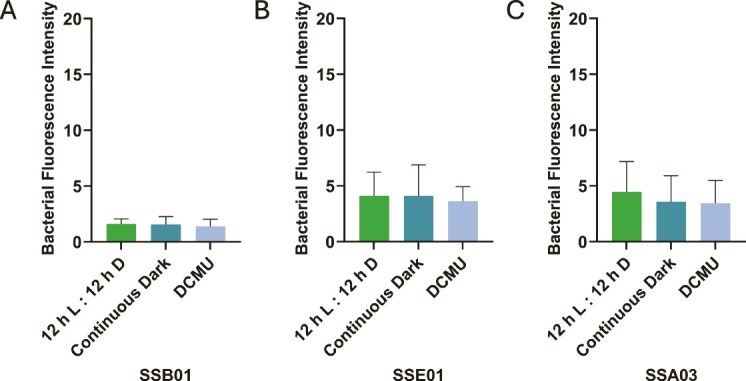
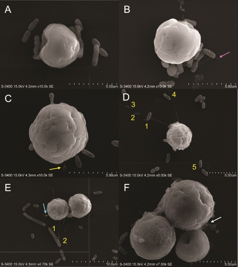
The cnidarian microbiome consists of a wide variety of beneficial microbes that play vital roles in maintaining and fortifying host health. Photosynthesis from symbiotic dinoflagellates (in the family Symbiodiniaceae) is crucial for their symbiosis establishment with the cnidarian host. Although more is known regarding interactions between the host and its associated bacteria and dinoflagellates, there has been little investigation into the relationship between the two microbes themselves and whether photosynthesis plays a role. Through two different methods of photosynthetic inhibition of dinoflagellates (incubation in the dark or pre-treatment with a photosystem II inhibitor), we investigated how pathogenic versus beneficial bacteria physically interact with three Symbiodiniaceae strains (symbiotic and free-living). The beneficial bacterium Tritonibacter mobilis appears to interact with photosynthesizing algae only. In the absence of photosynthesis, little to no physical interactions were observed between Symbiodiniaceae and T. mobilis. Bacterial congregation around individual dinoflagellate cells was significantly lower when photosynthesis was impaired, suggesting photosynthesis is a key facilitator of interactions between T. mobilis and all three Symbiodiniaceae strains. We also investigated whether photosynthesis affects interactions between Symbiodiniaceae and the pathogen Vibrio alginolyticus. Although no discernable impacts of photosynthetic inhibition were observed with the pathogen, scanning electron microscopy uncovered various mechanisms of interaction between Symbiodiniaceae and both bacteria, one of which includes the production of filaments not previously described. Overall, our research highlights the importance of photosynthesis in initiating interactions between bacteria and both free-living and symbiotic dinoflagellates, and opens a door to new questions regarding cell-surface interactions among individual microbes.

Keywords: algae; cnidarian; dinoflagellate; holobiont; microbiome; microscopy; symbiosis.


Conflict of interest statement

None declared.

Figures

Figure 1
Figure 1
Photosynthetic inhibition of Symbiodiniaceae reduces the localization of Tritonibacter mobilis (bacteria) to their algal-cell surfaces. Symbiodiniaceae strains SSB01 (A, B, C), SSE01 (D, E, F), and SSA03 (G, H, I) were pretreated for four days in a 12 h light:12 h dark cycle without DCMU (control; A, D, G), continuous dark cycle (B, E, H), or 12 h light:12 h dark cycle with 10 μM DCMU (C, F, I). IMK media was then replaced with sterile seawater (SSW) and GFP-tagged T. mobilis in SSW was introduced. Final concentrations of algae and bacteria in SSW were 2.0 × 105 cells ml−1 and OD600 = 0.1, respectively. Fluorescence imaging took place approximately 15 min after inoculation. Red, chlorophyll fluorescence of algae; green, recombinant GFP fluorescence of tagged T. mobilis. Scale bars represent 10 μm.
Figure 2
Figure 2
Inhibition of algal photosynthesis decreases Tritonibacter mobilis (bacteria) localization to Symbiodiniaceae. Algal strains (A) SSB01, (B) SSE01, and (C) SSA03 underwent the same three pretreatments: 12 h light:12 h dark cycle without DCMU (control), continuous dark cycle, or 12 h light:12 h dark cycle with 10 μM DCMU. Bacterial fluorescence (FITC channel) was measured in ImageJ. Sample sizes ranged from 29–34 algal cells for SSB01, 34–37 algal cells for SSE01, and 19–34 algal cells for SSA03, based on three independent trials of the experiment. Bars represent average bacterial fluorescence intensity + SD. A one-way ANOVA followed by a post-hoc Tukey’s test show significant differences where indicated. **P = .001. ns = not significant.
Figure 3
Figure 3
Scanning electron micrographs (SEM) of Tritonibacter mobilis (bacteria) physically interacting with Symbiodiniaceae strain SSB01. Algal cells were grown in IMK medium in a 12 h light:12 h dark cycle. Media was replaced with sterile seawater (SSW) and inoculated with T. mobilis in SSW. Final concentrations of algae and bacteria in SSW were 2.0 × 105 cells ml−1 and OD600 = 0.1, respectively. Algal–bacterial mixture was fixed approximately 15 min after inoculation for SEM (see materials and methods). (A, B) Star-shaped clusters of T. mobilis. (C–F) Specific cell–cell interactions between the polar ends of individual T. mobilis rods and SSB01. Leaf-shaped pads (arrows) at the ends of filamentous appendages protrude from the algal surface and interact with a bacterial rod.
Figure 4
Figure 4
SEM of Tritonibacter mobilis (bacteria) physically interacting with Symbiodiniaceae strain SSE01. Algal cells were grown in IMK medium in a 12 h light:12 h dark cycle. Media was replaced with sterile seawater (SSW) and inoculated with T. mobilis in SSW. Final concentrations of algae and bacteria in SSW were 2.0 × 105 cells ml−1 and OD600 = 0.1, respectively. Algal–bacterial mixture was fixed approximately 15 min after inoculation for SEM (see materials and methods). (A–E) Individual SSE01 cells interacting with T. mobilis. (F) Zoomed-out view of an SSE01 cluster with bacteria and surrounding extracellular matrix (ECM). ECM is labeled with arrows.
Figure 5
Figure 5
SEM of Tritonibacter mobilis (bacteria) physically interacting with Symbiodiniaceae strain SSA03. Algal cells were grown in IMK medium in a 12 h light:12 h dark cycle. Media was replaced with sterile seawater (SSW) and inoculated with T. mobilis in SSW. Final concentrations of algae and bacteria in SSW were 2.0 × 105 cells ml−1 and OD600 = 0.1, respectively. Algal–bacterial mixture was fixed approximately 15 min after inoculation for SEM (see materials and methods). (A) Magnified image of SSA03 tuft on cell surface next to and T. mobilis cluster. (B–E) Individual SSE01 cells interacting with T. mobilis. (F) Zoomed-out view of an SSE01 cluster with bacteria and ECM. ECM is labeled with arrows.
Figure 6
Figure 6
Photosynthetic inhibition of Symbiodiniaceae does not impact their interactions with Vibrio alginolyticus (bacteria). Symbiodiniaceae strains SSB01 (A, B, C), SSE01 (D, E, F), and SSA03 (G, H, I) were pretreated for four days in a 12 h light:12 h dark cycle without DCMU (control; A, D, G), continuous dark cycle (B, E, H), or 12 h light:12 h dark cycle with 10 μM DCMU (C, F, I). IMK media was then replaced with sterile seawater (SSW) and GFP-tagged V. alginolyticus in SSW was introduced. Final concentrations of algae and bacteria in SSW were 2.0 × 105 cells ml−1 and OD600 = 0.1, respectively. Fluorescence imaging took place approximately 15 min after inoculation. Red, chlorophyll fluorescence of algae; green, recombinant GFP fluorescence of tagged V. alginolyticus. Scale bars represent 10 μm.
Figure 7
Figure 7
Localization of Vibrio alginolyticus (bacteria) to Symbiodiniaceae is unchanged by photosynthetic inhibition. Algal strains (A) SSB01, (B) SSE01, and (C) SSA03 underwent the same three pretreatments: 12 h light:12 h dark cycle without DCMU (control), continuous dark cycle, or 12 h light:12 h dark cycle with 10 μM DCMU. Bacterial fluorescence (FITC channel) was measured in ImageJ. Sample sizes ranged from 22–39 algal cells for SSB01, 31–40 algal cells for SSE01, and 40–50 algal cells for SSA03, based on three independent trials of the experiment. Bars represent average bacterial fluorescence intensity + SD. A one-way ANOVA indicated no significant differences.
Figure 8
Figure 8
SEM of Vibrio alginolyticus (bacteria) physically interacting with Symbiodiniaceae strain SSB01. Algal cells were grown in IMK medium in a 12 h light:12 h dark cycle. Media was replaced with sterile seawater (SSW) and inoculated with T. mobilis in SSW. Final concentrations of algae and bacteria in SSW were 2.0 × 105 cells ml−1 and OD600 = 0.1, respectively. Algal–bacterial mixture was fixed approximately 15 min after inoculation for SEM (see materials and methods). (A, B) individual SSB01 cells interacting with V. alginolyticus with a bacterial flagellum present (pink arrow). (C) Individual SSB01 cell interacting with V. alginolyticus. SSB01 exhibits a small protruding filament attached to the poly-L-lysine-coated glass substratum (arrow). (D) SSB01 cell with multiple protruding filaments (numbers) which interact with either a bacterium or the glass substratum. (E) SSB01 cell with two protruding filaments (numbers) and two flagella branching from a peduncle (arrow). (F) Zoomed-out view of an SSB01 cluster covered in ECM (arrow) with surrounding bacteria.

Similar articles

References

    1. Yang X, Liu Z, Zhang Y et al. Dinoflagellate–bacteria interactions: physiology, ecology, and evolution. Biology 2024;13:579. 10.3390/biology13080579 - DOI - PMC - PubMed
    1. LaJeunesse TC, Parkinson JE, Gabrielson PW et al. Systematic revision of Symbiodiniaceae highlights the antiquity and diversity of coral endosymbionts. Curr Biol 2018;28:2570–2580.e6. 10.1016/j.cub.2018.07.008 - DOI - PubMed
    1. Tran C. Coral–microbe interactions: their importance to reef function and survival. Emerg Top Life Sci 2022;6:33–44. 10.1042/ETLS20210229 - DOI - PubMed
    1. Matthews JL, Raina J, Kahlke T et al. Symbiodiniaceae-bacteria interactions: rethinking metabolite exchange in reef-building corals as multi-partner metabolic networks. Environ Microbiol 2020;22:1675–87. 10.1111/1462-2920.14918 - DOI - PubMed
    1. Warner ME, Fitt WK, Schmidt GW. Damage to photosystem II in symbiotic dinoflagellates: a determinant of coral bleaching. Proc Natl Acad Sci 1999;96:8007–12. 10.1073/pnas.96.14.8007 - DOI - PMC - PubMed

LinkOut - more resources