. 2025 May 15;27(Suppl 5):v256-v279.
doi: 10.1093/eurheartjsupp/suaf077. eCollection 2025 May.

ANMCO position paper 'Hypertrophic cardiomyopathy: from diagnosis to treatment'

Affiliations

ANMCO position paper 'Hypertrophic cardiomyopathy: from diagnosis to treatment'

Cristina Chimenti et al. Eur Heart J Suppl. .

Abstract

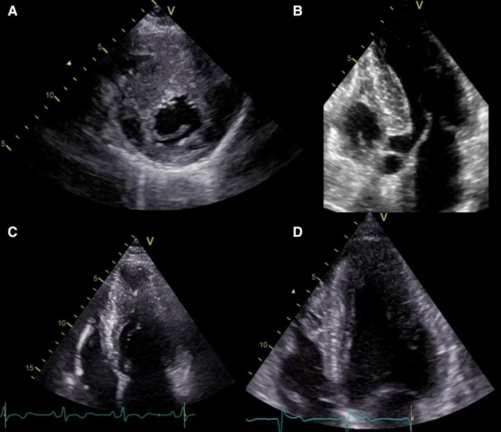
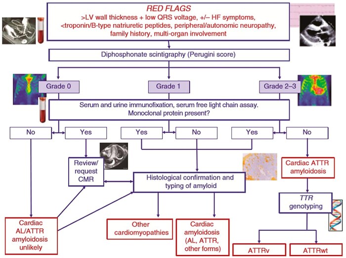
Hypertrophic cardiomyopathy (HCM) is a non-rare genetic cardiomyopathy, with an estimated prevalence of 1:500, characterized by an increase in the left ventricular wall thickness in the absence of increased loading conditions. The hypertrophy is mostly asymmetric and predominantly affects the basal septum and anterior wall. Left ventricular outflow tract obstruction, at rest or after provocative tests, is detected in many patients and represents the primary cause of reduced functional capacity, as well as an independent predictor of sudden cardiac death and advanced heart failure. Until ∼1 year ago, symptomatic patients despite maximal therapy with β-blockers or calcium channel blockers plus disopyramide had only basal septal reduction therapy through myectomy or septal alcoholization as additional therapeutic options. Today, a new class of drugs that inhibit cardiac myosin activity is available for patients with obstructive HCM. In light of the new treatment perspectives, the correct clinical-therapeutic classification of affected patients becomes of fundamental importance for the cardiologist. The aim of this position paper is to increase the knowledge of cardiologists in the field of HCM, defining its epidemiological, genetic, and pathological characteristics, identifying the diagnostic criteria and instrumental methods capable of stratifying the risk profile, with the aim of an optimal therapy tailored on the single patient.

Keywords: Diagnosis; Heart failure; Hypertrophic cardiomyopathy; Therapy; Ventricular hypertrophy.


Conflict of interest statement

Conflict of interest: none declared.

Figures

Graphical Abstract
Graphical Abstract
Integrated approach to hypertrophic cardiomyopathy, which includes genetic evaluation, family screening, morphological characterization, evaluation of disease signs and symptoms, electrocardiogram study, multimedia imaging, differential diagnosis with phenocopies, and personalized treatment.
Figure 1
Figure 1
Clinical-genetic course of hypertrophic cardiomyopathy. HCM, hypertrophic cardiomyopathy.
Figure 2
Figure 2
Hypertrophic cardiomyopathy: macroscopic pictures. (A) Asymmetrical septal form (case of sudden death). (B) Anterolateral asymmetric form (case of sudden death). (C) ‘End-stage’ form with reduced ejection fraction (case of death due to heart failure: diffuse intramyocardial fibrosis with thinning of the walls of the left ventricle). (D) ‘End-stage’ form with reduced ejection fraction (case of heart transplantation): diffuse intramyocardial fibrosis prevailing in the septum with thinning (bar = 20 mm in AD).
Figure 3
Figure 3
Hypertrophic cardiomyopathy: histological pictures. (A) Hypertrophy of myocytes and ‘disarray’ of individual cardiomyocytes (haematoxylin-eosin). (B) Fascicular ‘disarray’ with interstitial fibrosis (Azan Mallory trichrome). (C) Hypertrophy of the media with narrowing of the intramyocardial arteriole lumen (Azan Mallory trichrome). (D) Patches of replacement fibrosis in the context of cardiomyocyte ‘disarray’ (Azan Mallory trichrome).
Figure 4
Figure 4
Electrocardiographic red flags in the differential diagnosis of hypertrophic cardiomyopathy. AVB, atrioventricular block; RBBB, right bundle branch block; LVH, left ventricular hypertrophy; ST lat depr, lateral ST depression; Pre-exc ventr, ventricular pre-excitation; Neg Lat T, T negative in lateral leads; QT VT, QT vector discordance.
Figure 5
Figure 5
Different hypertrophic cardiomyopathy geometries at two-dimensional echocardiography: (A) Reverse septal curvature. (B) Sigmoid septum. (C) Mid-ventricular. (D) Apical.
Figure 6
Figure 6
Evaluation and treatment of left ventricular outflow tract obstruction in hypertrophic cardiomyopathy. Exercise echocardiography may be considered on an individual basis in presence of symptoms referable to latent obstruction and when the presence of a left ventricular outflow tract gradient is relevant to lifestyle advice and drug therapy decisions. *Valsalva manoeuvre, squatting, change from clinostatism to orthostatism. **Consider repeat examination after meal. HCM, hypertrophic cardiomyopathy.
Figure 7
Figure 7
Short-axis cardiac magnetic resonance imaging images of left ventricle in patient with history of malignant ventricular arrhythmias and sarcomeric hypertrophic cardiomyopathy. Left T2-weighted image with hyperintense areas of septal tissue oedema. In the centre same post-contrast image (LGE) with areas of myocardial fibrosis in the septal location that follow those of the T2-weighted image. Right quantification of areas of LGE (using 6 SD method) with extent of myocardial fibrosis estimated to be ∼15% of left ventricular mass. LGE, late gadolinium enhancement.
Figure 8
Figure 8
Arrhythmia management and prevention of sudden death. ESC, European Society of Cardiology; HCM, hypertrophic cardiomyopathy; ICD, implantable cardioverter defibrillator; LGE, late gadolinium enhancement; LVEF, left ventricular ejection fraction.
Figure 9
Figure 9
Comparison of flow chart of treatment in patients with hypertrophic obstructive cardiomyopathy. Adapted from European Society of Cardiology guidelines 2023 (2) and American Heart Association/American College of Cardiology 2024 (1). ACC, American College of Cardiology; AHA, American Heart Association; ESC, European Society of Cardiology; LVOTO, left ventricular outflow tract obstruction.
Figure 10
Figure 10
Pathogenesis of cardiac damage, main clinical manifestations, extracardiac and cardiac red flags and diagnostic pathway in Fabry disease cardiomyopathy.
Figure 11
Figure 11
Diagnostic–therapeutic course of cardiac amyloidosis. AL, light chain; ATTR, amyloidosis transthyretin; ATTRwt, wild-type amyloidosis transthyretin; CMR, cardiac magnetic resonance.

References

    1. Ommen SR, Ho CY, Asif IM, Balaji S, Burke MA, Day SM et al. 2024 AHA/ACC/AMSSM/HRS/PACES/SCMR guideline for the management of hypertrophic cardiomyopathy: a report of the American Heart Association/American College of Cardiology Joint Committee on Clinical Practice Guidelines. Circulation 2024;149:e1239–e1311. - PubMed
    1. Arbelo E, Protonotarios A, Gimeno JR, Arbustini E, Barriales-Villa R, Basso C et al. 2023 ESC guidelines for the management of cardiomyopathies. Eur Heart J 2023;44:3503–3626. - PubMed
    1. Bertero E, Canepa M, Pieroni M, Olivotto I. Update sulla gestione del paziente con cardiomiopatia ipertrofica alla luce delle nuove linee guida nord-americane. G Ital Cardiol (Rome) 2025;26:185–194. - PubMed
    1. Semsarian C, Ingles J, Maron MS, Maron BJ. New perspectives on the prevalence of hypertrophic cardiomyopathy. J Am Coll Cardiol 2015;65:1249–1254. - PubMed
    1. Maron BJ, Gardin JM, Flack JM, Gidding SS, Kurosaki TT, Bild DE. Prevalence of hypertrophic cardiomyopathy in a general population of young adults. Echocardiographic analysis of 4111 subjects in the CARDIA study. Coronary artery risk development in (young) adults. Circulation 1995;92:785–789. - PubMed

LinkOut - more resources