

Management of Periapical Lesion and Discoloration with Periapical Microsurgery Followed up by Internal-External Bleaching and Direct Composite Restoration: One-Year Clinical Evaluation
- PMID: 40385504
- PMCID: PMC12085892
- DOI: 10.2147/CCIDE.S516207
Management of Periapical Lesion and Discoloration with Periapical Microsurgery Followed up by Internal-External Bleaching and Direct Composite Restoration: One-Year Clinical Evaluation
Abstract
Background: Endodontic treatment outcomes may change over time. Factors such as compromised coronal seal and inadequate obturation can lead to canal reinfection and often periapical infection, thus developing into periapical lesions. Biofilm occupying the surface of the root tip can cause failure of conventional endodontic treatment or retreatment. Healing is achievable depending on the success of efforts to eradicate the biofilm layer from the site. Tooth discoloration after a root canal treatment can also be concerning, with several causes and treatment options. One of the minimally invasive aesthetic treatment options for discolored anterior teeth is dental bleaching or direct composite veneers.
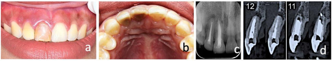
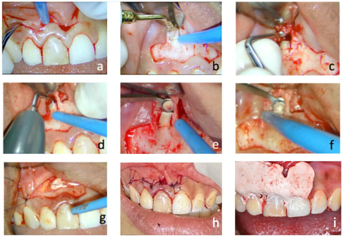
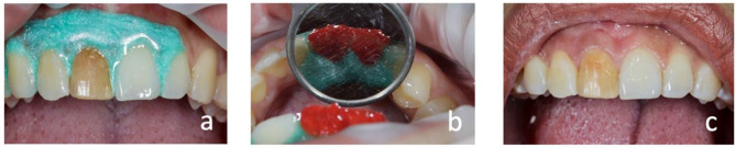
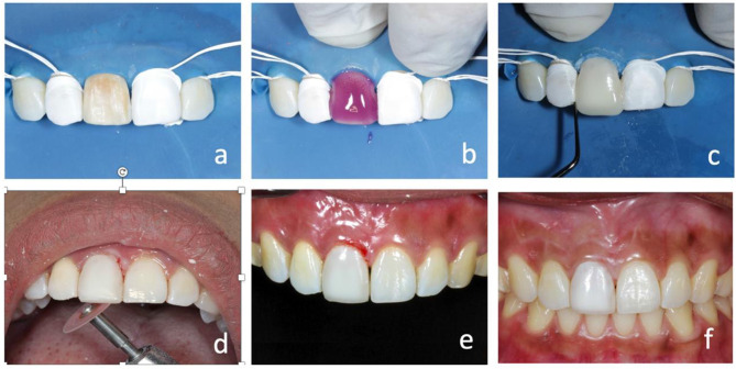
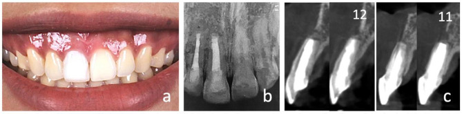
Case report: A 22-year-old patient experienced pain in the right maxillary lateral and central incisors with a history of trauma at the age of six. Both teeth have undergone root canal treatment at the age of ten years. Clinically, the teeth were discolored with visible old composite restorations. Radiological finding shows radiolucency in the periapical and compromised apical structure. Endodontic retreatment and periapical microsurgery were performed to remove the periapical lesion, followed by internal-external bleaching and direct composite veneer to restore the aesthetics.
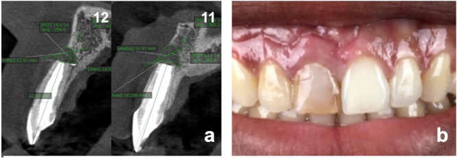
Results: The procedure was successful, and a one-year follow-up recall revealed bone regeneration around their apices and esthetic outcomes that satisfied the patient.
Conclusion: The technique described is an excellent approach in conserving the tooth with periapical lesions and compromised apical structure due to root resorption and internally stained teeth.
Keywords: direct composite veneer; internal-external bleaching; periapical lesion; periapical microsurgery; tooth discoloration.
© 2025 Hidayat et al.
Conflict of interest statement
The authors report no conflicts of interest in this work.
Figures
Similar articles
-
Endo-restorative treatment of a severly discolored upper incisor: resolution of the "aesthetic" problem through Componeer veneering System.Ann Stomatol (Roma). 2016 Feb 12;6(3-4):113-8. doi: 10.11138/ads/2015.6.3.113. eCollection 2015 Jul-Dec. Ann Stomatol (Roma). 2016. PMID: 26941900 Free PMC article. Review.
-
Healing beyond the apex: Nonsurgical management of a periapical lesion with esthetic rehabilitation using direct composite veneers.J Conserv Dent Endod. 2025 Jul;28(7):704-707. doi: 10.4103/JCDE.JCDE_244_25. Epub 2025 Jul 2. J Conserv Dent Endod. 2025. PMID: 40746477 Free PMC article.
-
Horizontal bone augmentation of the edentulous area with simultaneous endodontic microsurgery of the adjacent tooth: A digitally-driven multidisciplinary case report with a 1-year follow-up.Int J Oral Implantol (Berl). 2021 Nov 2;14(4):435-451. Int J Oral Implantol (Berl). 2021. PMID: 34726851
-
Persistent extraradicular infection in root-filled asymptomatic human tooth: scanning electron microscopic analysis and microbial investigation after apical microsurgery.J Endod. 2011 Dec;37(12):1696-700. doi: 10.1016/j.joen.2011.09.018. J Endod. 2011. PMID: 22099908
-
Limitations of previously published systematic reviews evaluating the outcome of endodontic treatment.Int Endod J. 2009 Aug;42(8):656-66. doi: 10.1111/j.1365-2591.2009.01600.x. Epub 2009 Jun 22. Int Endod J. 2009. PMID: 19548929 Review.
References
Publication types
LinkOut - more resources
Full Text Sources
