. 2025 Jan 23;10(3):e10748.
doi: 10.1002/btm2.10748. eCollection 2025 May.

Amelioration of biased neuronal differentiation in humanized mouse model of valproic acid-induced autism by precisely targeted transcranial magnetic stimulation

Affiliations

Amelioration of biased neuronal differentiation in humanized mouse model of valproic acid-induced autism by precisely targeted transcranial magnetic stimulation

Yilin Hou et al. Bioeng Transl Med. .

Abstract

Autism spectrum disorder (ASD) is a group of developmental diseases, which still lacks effective treatments. Pregnant exposure of Valproic acid (VPA) is an important environmental risk factor for ASD, but it's long-term effects on the development of human neural cells, particularly in vivo, and the corresponding treatment have yet been fully investigated. In the present study, we first made a humanized ASD mouse model by transplanting VPA-pretreated human neural progenitor cells (hNPCs) into the cortex of immune-deficient mice. In comparison with wild type and control chimeric mice, ASD chimeric mice (VPAhNPC mice) exhibit core syndromes of ASD, namely dramatic reduction of sociability, social interaction and social communication, and remarkable increase of stereotype repetitive behaviors and anxiety-like behaviors. At cellular level, VPA-pretreatment biased the differentiation of human excitatory neurons and their axonal projections in host brain. Chemogenetic suppression of human neuronal activity restored most behavior abnormalities of VPAhNPC mice. Further, specific modulation of human neurons by a newly developed transcranial magnetic stimulation (TMS) device which could precisely target hPNCs effectively recued the core syndromes of ASD-like behaviors, restored the excitatory-inhibitory neuronal differentiation and axonal projection, and reversed the expression of over half of the VPA-affected genes. These data demonstrated that VPAhNPC mice could be used as a humanized model of ASD and that precisely targeted TMS could ameliorate the VPA-biased human neuronal differentiation in vivo.

Keywords: autism spectrum disorder; humanized mouse; precisely targeted TMS; valproic acid.


Conflict of interest statement

The authors declare no conflict of interest.

Figures

FIGURE 1
FIGURE 1
Establishment of VPA‐pretreated human neuron chimeric mice (VPAhNPC mice). (a) Experimental design for neural induction, VPA treatment and cell transplantation. (b) Double‐immunostaining and quantification of Nestin/H8‐GFP in hNPCs 3 days following VPA treatment. (c) Double‐immunostaining and quantification of Ki67/H8‐GFP in hNPCs 3 days following VPA treatment. (d) Double‐immunostaining and quantification of CaMKII/H8‐GFP in hNPCs 3 days following VPA treatment. (e) Immunostaining of GFP in the sagittal sections of VPAhNPC mice (left panel) and double‐immunostaining of Tuj‐1/H8‐GFP in VPAhNPC mice (right panel). (f) Double‐immunostaining of c‐Fos/H8‐GFP in VPAhNPC mice following social stimulation. Notice the wide distribution of GFP‐positive human nerve fibers in mouse brain and the expression of c‐Fos in human neurons. N = 5 batches of cells in (b–d). Students' t test in “b–d”. *p < 0.05.**p < 0.01. Con, control.
FIGURE 2
FIGURE 2
Social behaviors of VPAhNPC mice. (a) Three‐chamber tests of social preference. (b) Three‐chamber tests of social novelty. Notice the decrease of social preference and social novelty in VPAhNPC mice. (c) Resident‐intruder assay. (d) Ultrasonic vocalization. Notice the decrease of social interaction and ultrasonic communication in VPAhNPC mice. N = 8 mice per group. One way ANOVA. *p < 0.05.**p < 0.01. ***p < 0.001. WT, wild type. Chimera, control hNPC chimeric mice. Chimera+VPA, VPAhNPC mice.
FIGURE 3
FIGURE 3
Repetitive and anxiety‐like behaviors of VPAhNPC mice. (a) Grooming test of WT mice, control chimeric mice and VPAhNPC mice. (b) Marble burying test. Notice the increase of grooming and marbles buried in VPAhNPC mice. (c) Elevated plus maze test of WT mice, control chimeric mice and VPAhNPC mice. (d) Open field test of WT mice, control chimeric mice and VPAhNPC mice. VPAhNPC mice showed reduced movement in the open arm and center field. N = 8 mice per group. One way ANOVA. *p < 0.05.**p < 0.01. Chimera, control chimeric mice; VPA, VPAhNPC mice; WT, wild type.
FIGURE 4
FIGURE 4
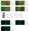
Biased excitatory neuronal differentiation and axonal projection of human neurons in VPAhNPC mice. (a) Double‐immunostaining of Tuj1/H8‐GFP in control chimeric mice and VPAhNPC mice, and quantification. (b) Double‐immunostaining of CaMKII/H8‐GFP in control chimeric mice and VPAhNPC mice, and quantification. (c) Double‐immunostaining of Vglut1/H8‐GFP in control chimeric mice and VPAhNPC mice, and quantification. (d) Double‐immunostaining of GABA/H8‐GFP in control chimeric mice and VPAhNPC mice, and quantification. Notice the enhanced generation of CaMKII‐positive and Vglut1‐positive neurons and decreased generation of GABA‐positive neurons from VPA‐pretreated hNPCs. (e) Western‐blotting and quantification of Tuj‐1, CamKII, vGlut1 and GAD‐67 in the hNPC graft of control chimeric mice and VPAhNPC mice. (f–h) GFP‐positive human axons in the mPFC, striatum and hippocampus of control chimeric mice and VPAhNPC mice. Notice that there were more human axons in the mPFC, striatum and hippocampus of VPAhNPC mice. N = 5 mice per group. Student's t test. *p < 0.05. **p < 0.01. Con, control chimeric mice; VPA, VPAhNPC mice.
FIGURE 5
FIGURE 5
Alleviation of social defects of VPAhNPC mice by chemogenetically inhibiting human excitatory neurons. (a) Experimental design for “b–e”. (b, c) 3‐chamber test of WT mice, VPA‐hM4DihNPC mice treated with vehicle or CNO. (d) Resident‐intruder assay of WT mice, VPA‐hM4DihNPC mice treated with vehicle or CNO. (e) Ultrasonic vocalization of WT mice, VPA‐hM4DihNPC mice treated with vehicle or CNO. Notice the improvement of social preference, social interaction and social communication of VPA‐hM4DihNPC mice by CNO. N = 8 mice per group. One way ANOVA in “b–e”. *p < 0.05.**p < 0.01. ***p < 0.001. CNO, clozapine‐N‐oxide; Con, control chimeric mice; VPA‐hM4Di, VPA‐hM4DihNPC mice.
FIGURE 6
FIGURE 6
Alleviation of repetitive behaviors of VPAhNPC mice by chemogenetically inhibiting human excitatory neurons. (a) Grooming test of WT mice, VPA‐hM4DihNPC mice treated with vehicle or CNO. (b) Marble burying test of WT mice, VPA‐hM4DihNPC mice treated with vehicle or CNO. Notice the decrease of repetitive behavior of VPA‐hM4DihNPC mice by CNO treatment. (c) Elevated plus maze test of WT mice, VPA‐hM4DihNPC mice treated with vehicle or CNO. (d) Open field test of WT mice, VPA‐hM4DihNPC mice treated with vehicle or CNO. Notice that CNO treatment significantly enhanced animal movement in the open arm and center field. N = 8 mice per group. One way ANOVA. *p < 0.05.**p < 0.01. ***p < 0.001. CNO, clozapine‐N‐oxide; Con, control chimeric mice; VPA‐hM4Di, VPA‐hM4DihNPC mice.
FIGURE 7
FIGURE 7
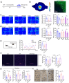
Amelioration of ASD‐like behaviors of VPAhNPC mice by precisely targeted rTMS. (a) Experimental design for “b–h”. (b) Computer simulation of the rTMS‐induced electric field and c‐Fos staining following rTMS treatment. Notice the c‐Fos‐positive cells in hNPC graft but not in mouse cortex. (c, d) Three‐chamber test of WT mice, VPAhNPC mice treated with or without rTMS. (e) Resident‐intruder assay of WT mice, VPAhNPC mice treated with or without rTMS. (f) Ultrasonic vocalization of WT mice, VPAhNPC mice treated with or without rTMS. Notice the increase of social preference, social interaction and ultrasonic communication of VPAhNPC mice by precisely targeted rTMS. (g, h) Grooming test and marble burying test of WT mice, VPAhNPC mice treated with or without rTMS. Notice the decrease of repetitive behavior of VPAhNPC mice by rTMS treatment. N = 10 mice per group. One way ANOVA. *p < 0.05.**p < 0.01. ***p < 0.001. Con, control chimeric mice; rTMS, repeated transcranial magnetic stimulation; VPA, VPAhNPC mice.
FIGURE 8
FIGURE 8
Restoration of neuronal differentiation and axonal projection of VPAhNPC mice by precisely targeted rTMS. (a) Double‐immunostaining of CaMKII/H8‐GFP in WT mice, VPAhNPC mice treated with or without rTMS. (b) Double‐immunostaining of Vglut1/H8‐GFP in WT mice, VPAhNPC mice treated with or without rTMS. (c) Double‐immunostaining of GABA/H8‐GFP in WT mice, VPAhNPC mice treated with or without rTMS. Notice the decrease of CaMKII/H8‐GFP‐positive and Vglut1/GFP‐positive cells, but the increase of GABA/H8‐GFP‐positive cells in rTMS‐treated VPAhNPC mice. (d–f) GFP‐staining in the mPFC, striatum and hippocampus. Notice the decrease of human axons in the mPFC and striatum of rTMS‐treated VPAhNPC mice. (g) Western‐blotting and quantification of Tuj‐1, CamKII, vGlut1 and GAD‐67 in the hNPC graft of control chimeric mice, VPAhNPC mice and rTMS‐treated VPAhNPC mice. (h–j) Heat‐map, Venn diagram and GO analysis of “up‐down” regulated genes in hNPCs from control chimeric mice, VPAhNPC mice and rTMS‐treated VPAhNPC mice. Notice the 157 “up‐down” genes and the enrichment of “neurogenesis” and “cell differentiation” in the top GO list. (k) qPCR validation of common top genes (6 out of 10) in “neurogenesis” and “cell differentiation”. N = 4–6 mice per group (a–g), 3 mice per group (h–k). One way ANOVA. *p < 0.05.**p < 0.01. ***p < 0.001. Con, control chimeric mice; rTMS, repeated transcranial magnetic stimulation; VPA, VPAhNPC mice.

Similar articles

References

    1. Manoli DS, State MW. Autism spectrum disorder genetics and the search for pathological mechanisms. Am J Psychiatry. 2021;178(1):30‐38. - PMC - PubMed
    1. Sohal VS, Rubenstein JLR. Excitation‐inhibition balance as a framework for investigating mechanisms in neuropsychiatric disorders. Mol Psychiatry. 2019;24(9):1248‐1257. - PMC - PubMed
    1. Legido A, Jethva R, Goldenthal MJ. Mitochondrial dysfunction in autism. Semin Pediatr Neurol. 2013;20(3):163‐175. - PubMed
    1. Thomas MS, Davis R, Karmiloff‐Smith A, Knowland VC, Charman T. The over‐pruning hypothesis of autism. Dev Sci. 2016;19(2):284‐305. - PubMed
    1. Jiang CC, Lin LS, Long S, et al. Signalling pathways in autism spectrum disorder: mechanisms and therapeutic implications. Signal Transduct Target Ther. 2022;7(1):229. - PMC - PubMed

LinkOut - more resources