. 2024 Oct 8;10(3):e10726.
doi: 10.1002/btm2.10726. eCollection 2025 May.

The hanging-heart chip: A portable microfluidic device for high-throughput generation of contractile embryonic stem cell-derived cardiac spheroids

Affiliations

The hanging-heart chip: A portable microfluidic device for high-throughput generation of contractile embryonic stem cell-derived cardiac spheroids

Pei-Tzu Lai et al. Bioeng Transl Med. .

Abstract

Stem cell-derived cardiac spheroids are promising models for cardiac research and drug testing. However, generating contracting cardiac spheroids remains challenging because of the laborious experimental procedure. Here, we present a microfluidic hanging-heart chip (HH-chip) that uses a microchannel and flow-driven system to facilitate cell loading and culture medium replacement operations to reduce the laborious manual handling involved in the generation of a large quantity of cardiac spheroids. The effectiveness of the HH-chip was demonstrated by simultaneously forming 50 mouse embryonic stem cell-derived embryonic bodies, which sequentially differentiated into 90% beating cardiac spheroids within 15 days of culture on the chip. A comparison of our HH-chip method with traditional hanging-drop and low-attachment plate methods revealed that the HH-chip could generate higher contracting proportions of cardiac spheroids with higher expression of cardiac markers. Additionally, we verified that the contraction frequencies of the cardiac spheroids generated from the HH-chip were sensitive to cardiotoxic drugs. Overall, our results suggest that the microfluidic hanging drop chip-based approach is a high-throughput and highly efficient method for generating contracting mouse embryonic stem cell-derived cardiac spheroids for cardiac toxicity and drug testing applications.

Keywords: cardiac spheroids; embryonic stem cell; hanging‐heart chip.


Conflict of interest statement

The authors declare that they have no conflict of interest.

Figures

FIGURE 1
FIGURE 1
Microfluidic hanging‐heart chip (HH‐chip) design. (a) Top and (b) side views of the channel structure. (c) Photograph showing droplets held by hydraulic pressure. (d) Top and (e) side views of the rack structure used with the microfluidic device. (f) Photograph of the device filled with color dye, placed within a 10 cm dish.
FIGURE 2
FIGURE 2
Operation procedure for the hanging‐heart chip. (a) Diagram of ES cell suspension loading into the channel at 3 × 104 cells/mL. (b) Diagram of cell sedimentation at the bottom of well. (c) Diagram showing the washing of cells on the channel structure. Scale bar = 1000 μm. (d) Diagram of the formation of EBs after 24 h incubation. Scale bar = 1000 μm. (e) Cells aggregate on microfluidic chip after 24 h. Scale bar = 500 μm. (f) Graph depicting the relationship between flow rate and the time required to inject the culture medium into the chip. Data are presented as the mean ± SD, n = 3.
FIGURE 3
FIGURE 3
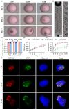
Cardiac spheroids generated on the heart‐hanging chip from mouse embryonic bodies using different cell densities. (a) Microscopy images showing EB morphology on ES culture on days 1, 2, and 7. Scale bar = 1000 μm. (b) Photograph of cell culture (initial cell load density = 3 × 104 cells/mL) 15 days post‐incubation on the chip. Scale bar = 4000 μm. (c) Graph illustrating the circularity of EBs at 24 and 48 h post‐seeding (n = 60, three independent experiments). (d) Graph illustrating the size of EBs 7 days post‐culture (n = 60, three independent experiments). (e) Graph illustrating the beating ratio from days 8–15 (n = 6, three independent experiments). (f) Confocal images of EBs. Alpha actinin 2‐positive and cardiac troponin T‐positive cells are stained red and green, respectively, and nuclei are stained with Hoechst (blue) (n = 6, three independent experiments). Scale bar = 200 μm. CTN2, alpha‐actinin 2; cTnT, cardiac troponin T. The asterisks indicate statistical significance, *p <0.05; **p <0.01; ***p <0.001; NS, nonsignificant; error bars show standard deviation.
FIGURE 4
FIGURE 4
Comparison of embryonic bodies formed using the heart‐hanging chip, traditional hanging‐drop, and low‐attachment 96‐well plate methods. (a) Bar graphs showing EB circularity at 24 and 48 h (n = 60, three independent experiments). (b) Graphs showing the change in EB size over a 7‐day culture period (n = 60, three independent experiments). (c) Graphs showing the beating ratio from days 8–15 (n = 6, three independent experiments). (d) Confocal images of EBs showing protein expression (n = 6, three independent experiments). Scale bar = 200 μm. (e) Bar graphs illustrating confocal image quantification of protein expression (n = 6, three independent experiments). For normalization, the intensities of ACTN2 and cTnT were normalized to the total cell number stained with Hoechst. Subsequently, the data were presented as fold changes, with the value of HH‐chip set to one‐fold. Data from the other two methods are indicated relative to this value. ACTN2, alpha‐actinin 2; cTnT, cardiac troponin T. The asterisks indicate statistical significance, *p <0.05; **p <0.01; ***p <0.001; NS: nonsignificant; NError bars show standard deviation.
FIGURE 5
FIGURE 5
Effects of drug‐induced cardiotoxicity on embryonic bodies formed using our hanging‐heart chip. (a) Graphs showing the frequency of beating of EBs with different patterns, and the frequency of beating EBs treated with 0, 0.01, and 1 μM isoproterenol for 10 min. (b) Quantification of the beating frequency changes before and after isoproterenol treatment. Data are presented as the mean ± SD, n = 6, in three independent experiments. (c) Microscopy images show morphology of EBs following exposure to 10, 30, and 50 μM doxorubicin. Scale bar = 500 μm. (d) Quantification of the spheroid size with doxorubicin treatment. Data are presented as the mean ± SD, n >15, in three independent experiments. (e) Quantification of cell viability with doxorubicin treatment. Data are presented as the mean ± SD, n = 9, in three independent experiments. (f) Graph showing the normalized beating frequency after exposure to 0–50 μM doxorubicin (n = 15, three independent experiments).

Similar articles

References

    1. Nguyen DC, Hookway TA, Wu Q, et al. Microscale generation of cardiospheres promotes robust enrichment of cardiomyocytes derived from human pluripotent stem cells. Stem Cell Reports. 2014;3:260‐268. doi:10.1016/j.stemcr.2014.06.002 - DOI - PMC - PubMed
    1. Jiang B, Xiang Z, Ai Z, et al. Generation of cardiac spheres from primate pluripotent stem cells in a small molecule‐based 3D system. Biomaterials. 2015;65:103‐114. doi:10.1016/j.biomaterials.2015.06.024 - DOI - PubMed
    1. Zuppinger C. 3D culture for cardiac cells. Biochim Biophys Acta. 2016;1863:1873‐1881. doi:10.1016/j.bbamcr.2015.11.036 - DOI - PubMed
    1. Kokkinopoulos I et al. Cardiomyocyte differentiation from mouse embryonic stem cells using a simple and defined protocol. Dev Dyn. 2016;245:157‐165. doi:10.1002/dvdy.24366 - DOI - PubMed
    1. Mercola M, Ruiz‐Lozano P, Schneider MD. Cardiac muscle regeneration: lessons from development. Genes Dev. 2011;25:299‐309. doi:10.1101/gad.2018411 - DOI - PMC - PubMed

LinkOut - more resources