

Size- and polymer-dependent toxicity of amorphous environmentally relevant micro- and nanoplastics in human bronchial epithelial cells
- PMID: 40385552
- PMCID: PMC12081513
- DOI: 10.1186/s43591-025-00126-9
Size- and polymer-dependent toxicity of amorphous environmentally relevant micro- and nanoplastics in human bronchial epithelial cells
Abstract
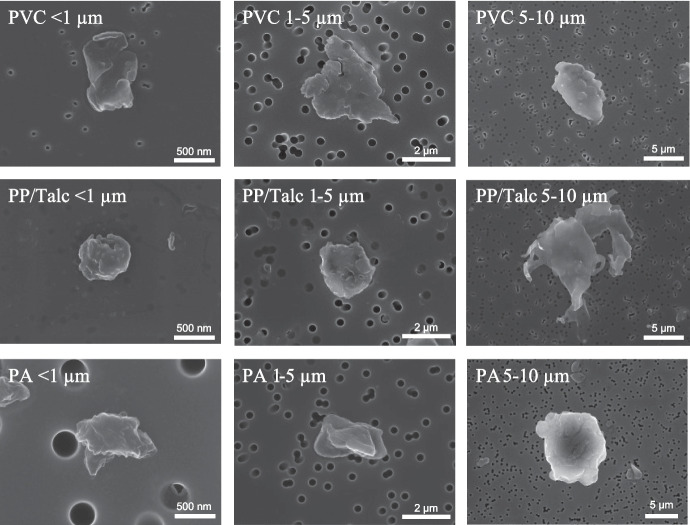
Background: Knowledge of the toxicological impact of micro- and nanoplastics (MNPs) on the human airway epithelium is limited and almost exclusively based on experiments applying high doses of spherical polystyrene (PS) particles. In this study, we investigated the toxicity of a broad size range of amorphous MNPs generated from different environmentally-relevant polymers.
Methods: Bronchial epithelial cells (BEAS-2B) were exposed to three different doses of polyvinylchloride (PVC), polypropylene (PP), or polyamide (PA) particles (< 1 μm-10 μm), as well as leachates from these polymers. Toxicity was evaluated by assessment of cytotoxicity, inflammation (IL-8 release and inflammatory gene expression) and oxidative stress (DCFH-DA assay and antioxidant gene expression). Furthermore, the molecular mechanism behind MNP-induced inflammation was investigated by studying activation of two well-known inflammation related transcriptional factors (NF-κB and AP-1).
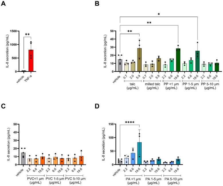
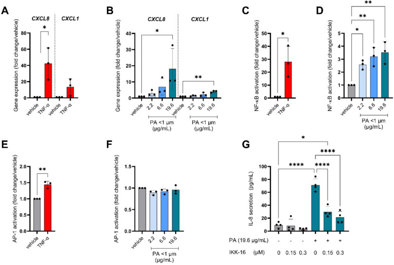
Results: Only PA nanoplastics induced significant cell death, IL-8 secretion and inflammatory gene expression compared to vehicle control. PA-induced inflammation was accompanied by NF-κB, but not AP-1, transcriptional activity. PA did not increase cellular ROS levels; however, it did lead to increased expression of the antioxidant gene superoxide dismutase 2. In addition to PA, exposure to < 1 µm and 1-5 µm PP particles resulted in elevated IL-8 secretion, likely due to the presence of talc added as filler. None of the leachates affected cytotoxicity or inflammation.
Conclusion: Toxicity of MNPs to human bronchial epithelial cells was dependent on polymer type, size and dose. Nanoplastics, especially PA, were more toxic to bronchial epithelial cells than microplastics and induced cytotoxicity and an inflammatory response.
Supplementary information: The online version contains supplementary material available at 10.1186/s43591-025-00126-9.
Keywords: Inflammation; Inhalation toxicology; NF-ĸB; Polyamide; Polypropylene; Polyvinylchloride.
© The Author(s) 2025.
Conflict of interest statement
Competing interestsThe authors declare no competing interests.
Figures


Similar articles
-
Assessing toxicity of amorphous nanoplastics in airway- and lung epithelial cells using air-liquid interface models.Chemosphere. 2024 Nov;368:143702. doi: 10.1016/j.chemosphere.2024.143702. Epub 2024 Nov 15. Chemosphere. 2024. PMID: 39522701
-
Micro- and nanoplastics (MNPs) and their potential toxicological outcomes: State of science, knowledge gaps and research needs.NanoImpact. 2023 Oct;32:100481. doi: 10.1016/j.impact.2023.100481. Epub 2023 Sep 16. NanoImpact. 2023. PMID: 37717636 Free PMC article. Review.
-
Understanding the Early Biological Effects of Isoprene-Derived Particulate Matter Enhanced by Anthropogenic Pollutants.Res Rep Health Eff Inst. 2019 Mar;2019(198):1-54. Res Rep Health Eff Inst. 2019. PMID: 31872748 Free PMC article.
-
In vitro effects of aged low-density polyethylene micro(nano)plastic particles on human airway epithelial cells.Environ Pollut. 2025 Jun 1;374:126186. doi: 10.1016/j.envpol.2025.126186. Epub 2025 Apr 2. Environ Pollut. 2025. PMID: 40185180
-
Micro- and nanoplastic toxicity: A review on size, type, source, and test-organism implications.Sci Total Environ. 2023 Jun 20;878:162954. doi: 10.1016/j.scitotenv.2023.162954. Epub 2023 Mar 21. Sci Total Environ. 2023. PMID: 36948318 Review.
References
-
- Prata JC. Airborne microplastics: consequences to human health? Environ Pollut. 2018;234:115–26. - PubMed
-
- Zhao X, Zhou Y, Liang C, Song J, Yu S, Liao G, et al. Airborne microplastics: Occurrence, sources, fate, risks and mitigation. Sci Total Environ. 2023;858:159943. - PubMed
-
- Dris R, Gasperi J, Rocher V, Saad M, Renault N, Tassin B. Microplastic contamination in an urban area: a case study in Greater Paris. Environ Chem. 2015;12(5):592–9.
-
- Jenner LC, Rotchell JM, Bennett RT, Cowen M, Tentzeris V, Sadofsky LR. Detection of microplastics in human lung tissue using μFTIR spectroscopy. Sci Total Environ. 2022;831:154907. - PubMed
LinkOut - more resources
Full Text Sources
Miscellaneous