. 2025 May 2:12:1526772.
doi: 10.3389/fmed.2025.1526772. eCollection 2025.

CD73+CD8+ T cells define a subset with anti-tumor potential in DLBCL patients

Affiliations

CD73+CD8+ T cells define a subset with anti-tumor potential in DLBCL patients

Lingyu Zhang et al. Front Med (Lausanne). .

Abstract

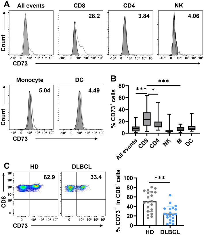
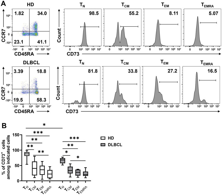
Introduction: CD73, a recently discovered immune checkpoint, catalyzes the conversion of AMP to adenosine, thereby suppressing anti-tumor immune responses.CD8+ T cells play a critical role in the immune response against cancer, yet their functionality can be modulated by various factors within the tumor microenvironment. In this study, we focus on identifying and characterizing CD73+CD8+ T cells in the peripheral blood of patients with diffuse large B-cell lymphoma (DLBCL), aiming to elucidate their functional and phenotypic roles in tumor immunity.

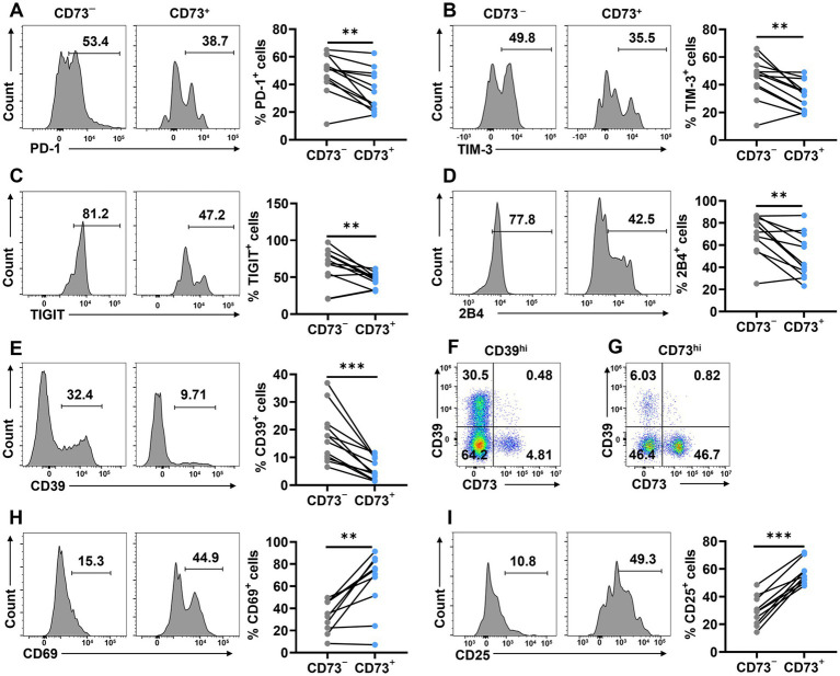
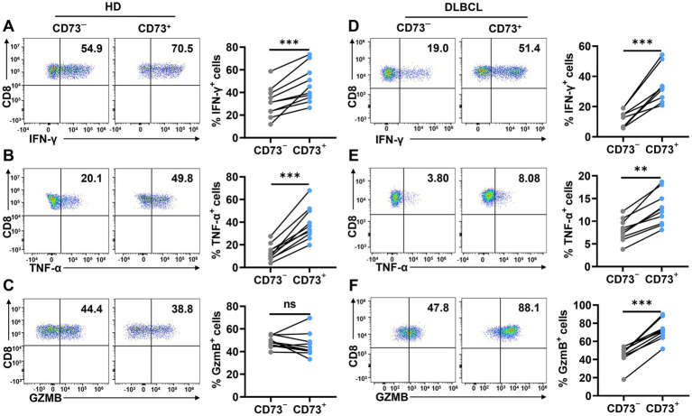
Methods: Using flow cytometry, we analyzed the expression of inhibitory receptors (e.g., PD-1, TIM-3) and activating markers (e.g., CD25, CD69) on CD73+CD8+ T cells compared to CD73-CD8+ T cells. In vitro functional assays were conducted to assess their cytotoxic activity against tumor cells, including cytokine production and tumor cell killing capacity.

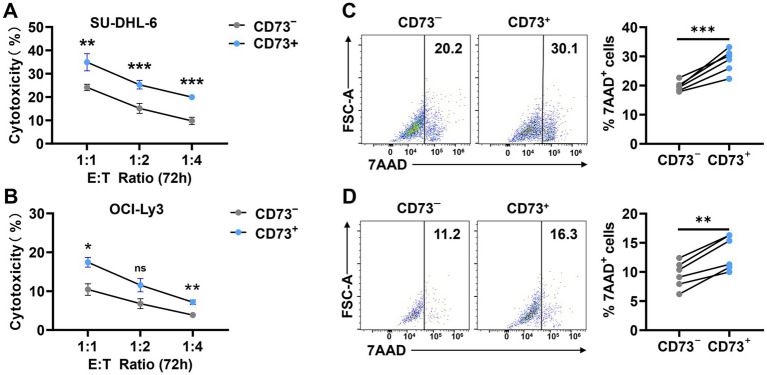
Results: CD73+CD8+ T cells exhibited a distinct immunophenotypic profile, characterized by reduced expression of inhibitory receptors and enhanced cytotoxic activity compared to their CD73- counterparts. These cells demonstrated higher levels of effector molecules (e.g., IFN-γ, TNF-α) and lower exhausted markers. The findings suggest that CD73+CD8+ T cells may retain stronger anti-tumor potential.

Discussion: This study highlights CD73+CD8+ T cells as a unique functional subset with potential therapeutic relevance in DLBCL. Their reduced exhaustion and heightened cytotoxicity position them as promising targets for immunotherapy strategies. However, the dual role of CD73 in adenosine-mediated immunosuppression warrants further investigation to reconcile its pro-tumorigenic effects with the observed anti-tumor activity of CD73+CD8+ T cells. Our findings deepen the understanding of CD8+ T cell heterogeneity in DLBCL and emphasize the need for mechanistic studies to explore CD73's context-dependent functions.

Keywords: CD73; CD8+ T cells; DLBCL; anti-tumor potential; cytotoxicity; immunotherapy.


Conflict of interest statement

The authors declare that the research was conducted in the absence of any commercial or financial relationships that could be construed as a potential conflict of interest.

Figures

Figure 1
Figure 1
CD73 is downregulated on CD8+ T cells in patients with DLBCL compared with HDs. (A,B) The expression of CD73 on all events, T cells (CD4+ T cells, CD8+ T cells), natural killer cells (NK), and myeloid cells (monocytes and dendritic cells) was assessed by flow cytometry. Representative flow data (A) and summary plot (B) of CD73. Data are shown as means ± standard deviation (SD). p values were obtained by the unpaired t test or Mann–Whitney test. *p < 0.05, ***p < 0.001. (C) Proportion of CD73+CD8+ T cells in total CD8+ T cells in HDs (n = 20) or patients with DLBCL (n = 20), each dot indicates one patient. Data are shown as means ± SD. p values were obtained by two-tailed unpaired t test. *p < 0.05, ***p < 0.001.
Figure 2
Figure 2
CD73 is less expressed on TEMRA. Expression of CD73 among each subset (TN, TCM, TEM, and TEMRA) of CD8 T cells was analyzed. Representative flow data (A) and plots (B) of percentage of CD73 expression among each subset of CD8+ T cells from HDs (n = 10) or DLBCL patients (n = 10) are shown. Data are shown as means ± SD. p values were obtained by Kruskal-Wallis test followed by Dunn’s multiple comparisons test, *p < 0.05; **p < 0.001; ***p < 0.001.
Figure 3
Figure 3
CD73+CD8+ T cells express fewer inhibitory and more activating receptors. (A–E) Proportion of PD-1+ (A), TIM-3+ (B), TIGIT+ (C),2B4+ (D) and CD39+ (E) cells in CD73+CD8+ T cells and CD73CD8+ T cells (n = 12). (F) Representative flow data of CD8+ T cells highly expressing CD39. (G) Representative flow data of CD8+ T cells highly expressing CD73. (H–I) Proportion of CD69+ (H), and CD25+ (I) cells in CD73+CD8+ T cells and CD73CD8+ T cells (n = 10). Data are the mean ± SD. A two-tailed paired t-test was used for statistical analyses, **p < 0.01, ***p < 0.001.
Figure 4
Figure 4
CD73+CD8+ T cells have higher effector functions. (A–C) Proportion of IFN-γ+ (A), TNF-α+ (B) and GzmB+ (C) cells in CD73+CD8+ T cells and CD73CD8+ T cells in HDs after stimulation with anti-CD3 (2 μg/ mL) and anti-CD28 (1 μg/mL) for 48 h (n = 10). (D–F) Proportion of IFN-γ+ (D), TNF-α+ (E) and GzmB+ (F) cells in CD73+CD8+ T cells and CD73CD8+ T cells in DLBCL after stimulation with anti-CD3 (2 μg/ mL) and anti-CD28 (1 μg/mL) for 48 h (n = 10). Data are the mean ± SD. A two-tailed paired t-test was used for statistical analyses, ns = non-significant, **p < 0.01, ***p < 0.001.
Figure 5
Figure 5
CD73+CD8+ T cells performing better anti-tumour effects. (A,B) Proportion of cytotoxicity after 72-h co-culture of CD73+CD8+ T cells or CD73CD8+ T cells with SU-DHL-6 (A) (n = 4) or OCI-Ly3 (B) (n = 4) cells at different effector-to-target (E: T = 1:1, 1:2, and 1:4) ratios. Data are shown as mean ± standard error of the mean (SEM). p values were obtained by two-tailed unpaired t test was used for statistical analyses, ns = non-significant, *p < 0.05; **p < 0.001; ***p < 0.001. (C,D) Representative flow data and plots of percentage of apoptosis after 72-h co-culture of CD73+CD8+ T cells or CD73CD8+ T cells with SU-DHL-6 (C) (n = 6) or OCI-Ly3 (D) (n = 6) cells. Data are the mean ± SD. p values were obtained by two-tailed paired t-test was used for statistical analyses, **p < 0.001; ***p < 0.001.

Similar articles

References

    1. Sassolas B, Labouche A, Guillet G. Herpes simplex virus infection associated with bullous pemphigoid. Br J Dermatol. (1993) 128:358–9. doi: 10.1111/j.1365-2133.1993.tb00187.x, PMID: - DOI - PubMed
    1. Li S, Young KH, Medeiros LJ. Diffuse large B-cell lymphoma. Pathology. (2018) 50:74–87. doi: 10.1016/j.pathol.2017.09.006, PMID: - DOI - PubMed
    1. Crump M, Neelapu SS, Farooq U, van den Neste E, Kuruvilla J, Westin J, et al. . Outcomes in refractory diffuse large B-cell lymphoma: results from the international SCHOLAR-1 study. Blood. (2017) 130:1800–8. doi: 10.1182/blood-2017-03-769620, PMID: - DOI - PMC - PubMed
    1. Carpio C, Bouabdallah R, Ysebaert L, Sancho JM, Salles G, Cordoba R, et al. . Avadomide monotherapy in relapsed/refractory DLBCL: safety, efficacy, and a predictive gene classifier. Blood. (2020) 135:996–1007. doi: 10.1182/blood.2019002395, PMID: - DOI - PMC - PubMed
    1. Philip M, Schietinger A. CD8+ T cell differentiation and dysfunction in cancer. Nat Rev Immunol. (2022) 22:209–23. doi: 10.1038/s41577-021-00574-3, PMID: - DOI - PMC - PubMed

LinkOut - more resources