A Study of Visual Outcomes and Spectral Domain Optical Coherence Tomography (SD-OCT) Biomarker Changes in Patients Treated With Ranibizumab for Diabetic Macular Edema in a Tertiary Hospital
- PMID: 40385737
- PMCID: PMC12085908
- DOI: 10.7759/cureus.82520
A Study of Visual Outcomes and Spectral Domain Optical Coherence Tomography (SD-OCT) Biomarker Changes in Patients Treated With Ranibizumab for Diabetic Macular Edema in a Tertiary Hospital
Abstract
Introduction: Diabetic macular edema (DME) is a leading cause of vision impairment in diabetes mellitus. Spectral-domain optical coherence tomography (SD-OCT) provides valuable biomarkers for assessing disease severity and treatment response. This study evaluates the visual outcomes and SD-OCT biomarker changes in patients treated with intravitreal ranibizumab at a tertiary hospital.
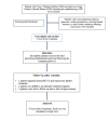
Materials and methods: A prospective cohort study was conducted on 50 Type 2 diabetes mellitus patients with center-involving DME at Employees' State Insurance Corporation (ESIC) Medical College and Hospital, Hyderabad, from July 2022 to December 2023. Inclusion criteria were best-corrected visual acuity (BCVA) <6/9 and central retinal thickness (CRT) ≥280 µm. Exclusion criteria included other retinal diseases, vision-impairing cataracts, or glaucoma. Patients received three monthly intravitreal ranibizumab injections and were followed up at one, two, three, and six months. BCVA, CRT, and SD-OCT biomarkers such as hyperreflective foci (HRF), subretinal neuroretinal detachment (SND), intraretinal cyst (IRC) size, disorganization of retinal inner layers (DRIL), ellipsoid zone (EZ) disruption, and external limiting membrane (ELM) disruption were assessed. Statistical analysis was performed using SPSS software, version 26 (IBM Corp., Armonk, NY).
Results: BCVA improved from 0.76±0.39 at baseline to 0.37±0.29 at six months (p<0.00001). CRT reduced from 473.66±111.65 µm to 326.64±71.37 µm (p<0.00001). HRF, SND, and IRC size showed significant regression. DRIL, EZ, and ELM disruption improved significantly (p<0.00001). There was no statistically significant association between OCT biomarker features and BCVA improvement of more than three lines.
Conclusion: Intravitreal ranibizumab significantly improves visual acuity and reduces retinal thickness in DME patients. SD-OCT biomarkers are valuable in monitoring treatment response and disease progression.
Keywords: diabetic macular edema; ranibizumab; retinal biomarkers; spectral-domain oct; visual acuity.
Copyright © 2025, Somagani et al.
Conflict of interest statement
Human subjects: Consent for treatment and open access publication was obtained or waived by all participants in this study. Institutional Ethics Committee, Employee's State Insurance Corporation (ESIC) Medical College and Hospital, Hyderabad issued approval 799/U/IEC/ESICMC/S0167/07/2022. Animal subjects: All authors have confirmed that this study did not involve animal subjects or tissue. Conflicts of interest: In compliance with the ICMJE uniform disclosure form, all authors declare the following: Payment/services info: All authors have declared that no financial support was received from any organization for the submitted work. Financial relationships: All authors have declared that they have no financial relationships at present or within the previous three years with any organizations that might have an interest in the submitted work. Other relationships: All authors have declared that there are no other relationships or activities that could appear to have influenced the submitted work.
Similar articles
-
Spectral-Domain OCT Predictors of Visual Outcomes after Ranibizumab Treatment for Macular Edema Resulting from Retinal Vein Occlusion.Ophthalmol Retina. 2020 Jan;4(1):67-76. doi: 10.1016/j.oret.2019.08.009. Epub 2019 Aug 28. Ophthalmol Retina. 2020. PMID: 31669329 Free PMC article. Clinical Trial.
-
Optical Coherence Tomography Biomarkers in Predicting Treatment Outcomes of Diabetic Macular Edema after Ranibizumab Injections.Medicina (Kaunas). 2023 Mar 21;59(3):629. doi: 10.3390/medicina59030629. Medicina (Kaunas). 2023. PMID: 36984630 Free PMC article.
-
Intravitreal Ranibizumab and Dexamethasone Implant Injections as Primary Treatment of Diabetic Macular Edema: The Month 24 Results from Simultaneously Double Protocol.Curr Eye Res. 2023 May;48(5):498-505. doi: 10.1080/02713683.2023.2168013. Epub 2023 Jan 12. Curr Eye Res. 2023. PMID: 36629472 Clinical Trial.
-
Practical Lessons from Protocol I for the Management of Diabetic Macular Edema.Dev Ophthalmol. 2017;60:91-108. doi: 10.1159/000459692. Epub 2017 Apr 20. Dev Ophthalmol. 2017. PMID: 28427069 Review.
-
Retinal Hyperreflective Foci Are Biomarkers of Ocular Disease: A Scoping Review With Evidence From Humans and Insights From Animal Models.J Ophthalmol. 2025 May 29;2025:9573587. doi: 10.1155/joph/9573587. eCollection 2025. J Ophthalmol. 2025. PMID: 40474962 Free PMC article. Review.
References
LinkOut - more resources
Full Text Sources
Research Materials

