Case Reports
. 2025 Apr 17;17(4):e82462.
doi: 10.7759/cureus.82462. eCollection 2025 Apr.

Total Knee Arthroplasty for Severe Crystalline-Induced Arthropathy: A Case Report From a Third-Level Hospital

Affiliations
Case Reports

Total Knee Arthroplasty for Severe Crystalline-Induced Arthropathy: A Case Report From a Third-Level Hospital

Diego Hernández-Penagos et al. Cureus. .

Abstract

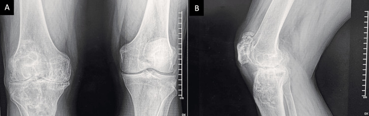
Gout is an inflammatory arthritis caused by monosodium urate crystal deposition, leading to progressive joint destruction and functional impairment. While pharmacologic treatment remains the standard, advanced cases with intra-articular and intraosseous tophi may require surgical intervention. Total knee arthroplasty (TKA) has been reported as a viable option for managing severe tophaceous gout with structural bone defects, improving joint function and pain control when conservative therapy fails. We present the case of a 56-year-old male with a long-standing history of gout and progressive knee pain refractory to medical treatment. Imaging revealed extensive intraosseous tophi with cavitary bone defects, prompting the decision for TKA with bone allograft reconstruction. The patient experienced favorable postoperative recovery, demonstrating significant improvement in joint mobility and function, with no complications observed. This case highlights the role of surgical intervention in the management of advanced tophaceous gout as part of an integrated approach alongside pharmacologic control.

Keywords: arthroplasty; gout disease; joint replacement surgeon; knee arthroplasty; knee replaecment.


Conflict of interest statement

Human subjects: Consent for treatment and open access publication was obtained or waived by all participants in this study. Hospital Regional Elvia Carrillo Puerto, Institute for Social Security and Services for State Workers (ISSSTE) issued approval RPI/HRECP/078. Conflicts of interest: In compliance with the ICMJE uniform disclosure form, all authors declare the following: Payment/services info: All authors have declared that no financial support was received from any organization for the submitted work. Financial relationships: All authors have declared that they have no financial relationships at present or within the previous three years with any organizations that might have an interest in the submitted work. Other relationships: All authors have declared that there are no other relationships or activities that could appear to have influenced the submitted work.

Figures

Figure 1
Figure 1. Preoperative range of motion of the right knee
A: maximum flexion of the right knee in lateral view: 85º; B: maximum extension of the right knee, showing a flexion contracture of 30º.
Figure 2
Figure 2. Radiographic images of the right knee
A: anteroposterior; B: lateral projections show a bilateral reduction in joint space with varus deformity. Marginal lytic bone lesions are noted at the subchondral level of the proximal tibia and distal femur, exhibiting well-defined sclerotic edges and no periosteal reaction. At the patellar level, there is a reduction in joint space along with marginal osteophytes present at the upper pole.
Figure 3
Figure 3. Intraoperative photograph of the right knee
A: significant infiltration of the femoral articular cartilage (white arrow) by tophaceous urate deposits is observed, along with a cavitary bone lesion in the subchondral bone of the proximal right tibia (black arrow) filled with semi-solid yellowish tissue; B: cavitary bone lesion in the subchondral region of the right lateral condyle prior to anterior and posterior femoral cuts (black arrow); C: full extent, depth, and diameter of the cavitary lesion in the subchondral bone of the proximal tibia and distal femur (black arrow); D: filling of the cavitary bone defect in the proximal tibia with bone allograft chips and preparation for the placement of prosthetic implants (black arrow).
Figure 4
Figure 4. Intraoperative photograph of the angular correction and range of motion improvement
A: maximum flexion of the right knee in lateral view: 110º; B: maximum extension of the right knee: 0º.
Figure 5
Figure 5. Clinical photograph of angular correction and range of motion improvement six weeks post-surgery
A: maximum flexion of the right knee in lateral view: 110º; B: maximum extension of the right knee: 0º, and close surgical wound after six weeks post-surgery.
Figure 6
Figure 6. Postoperative X-ray of the right knee joint
A: anteroposterior; B: lateral view, and the bone allograft appeared to be intact with minimal remodeling.

Similar articles

References

    1. Management and cure of gouty arthritis. Keller SF, Mandell BF. Med Clin North Am. 2021;105:297–310. - PubMed
    1. Unusual case of pseudotumoral hip injury due to gout: case report (Article in Spanish) Narváez-Carrasquilla G, Paternina-Carballo JD, Narváez-Rodríguez G. Rev Med Inst Mex Seguro Soc. 2023;61:895–899. - PMC - PubMed
    1. Gout after total knee arthroplasty. Chernoff DJ, Barker JP, Wingerter SA, Shriwise TL. Arthroplast Today. 2020;6:278–282. - PMC - PubMed
    1. Clinics in diagnostic imaging (200). Intra-articular chronic tophaceous gout of the knee. Lee YW, Kok SX, Wong LW, Wong BS. Singapore Med J. 2019;60:502–507. - PMC - PubMed
    1. Pseudogout in the early postoperative period after total knee arthroplasty. Harato K, Yoshida H. J Arthroplasty. 2013;28:374. - PubMed

Publication types

LinkOut - more resources