

Targeted Muscle Reinnervation-an Up-to-Date Review: Evidence, Indications, and Technique
- PMID: 40386007
- PMCID: PMC12081100
- DOI: 10.1055/a-2521-2199
Targeted Muscle Reinnervation-an Up-to-Date Review: Evidence, Indications, and Technique
Abstract
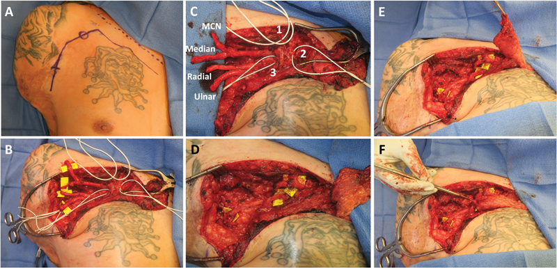
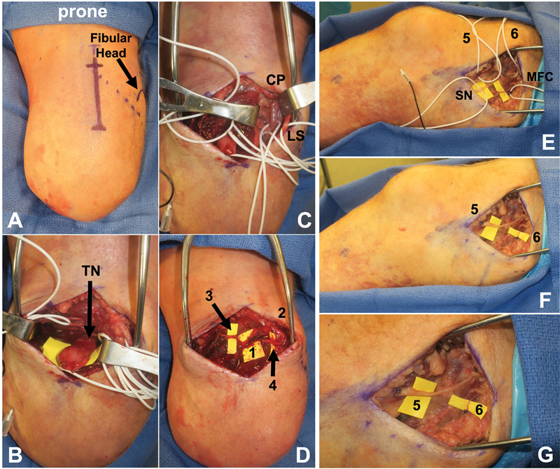
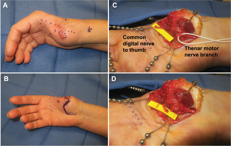
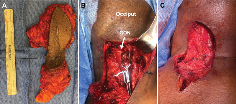
Targeted muscle reinnervation (TMR) is a surgical technique originally created to improve prosthetic function following upper extremity amputation. TMR has since been shown to be effective in the prevention and treatment of chronic postamputation phantom and residual limb pain in both upper and lower extremity amputees and for neurogenic pain in the nonamputee patient population. This article provides a current review of the various indications for TMR and surgical techniques, organized by amputation site, timing, and regional anatomy.
Keywords: myoelectric prosthesis; nerve transfers; painful neuromas; targeted muscle reinnervation.
The Author(s). This is an open access article published by Thieme under the terms of the Creative Commons Attribution License, permitting unrestricted use, distribution, and reproduction so long as the original work is properly cited. ( https://creativecommons.org/licenses/by/4.0/ ).
Conflict of interest statement
Conflict of Interest G.A.D. and J.H.K. are consultants for Checkpoint Surgical, Inc. A.G.C., M.D.R., and S.P. have no disclosures.
Figures
Similar articles
-
Targeted Muscle Reinnervation and Regenerative Peripheral Nerve Interface for Myoelectric Prosthesis Control: The State of Evidence.Ann Plast Surg. 2025 Jun 1;94(6S Suppl 4):S572-S576. doi: 10.1097/SAP.0000000000004273. Ann Plast Surg. 2025. PMID: 40459463 Review.
-
Targeted muscle reinnervation: a novel approach to postamputation neuroma pain.Clin Orthop Relat Res. 2014 Oct;472(10):2984-90. doi: 10.1007/s11999-014-3528-7. Clin Orthop Relat Res. 2014. PMID: 24562875 Free PMC article.
-
Targeted Muscle Reinnervation in the Hand: Treatment and Prevention of Pain After Ray Amputation.J Hand Surg Am. 2020 Sep;45(9):884.e1-884.e6. doi: 10.1016/j.jhsa.2019.10.020. Epub 2019 Dec 6. J Hand Surg Am. 2020. PMID: 31818541
-
Targeted Muscle Reinnervation: A Paradigm Shift for Neuroma Management and Improved Prosthesis Control in Major Limb Amputees.J Am Acad Orthop Surg. 2021 Apr 1;29(7):288-296. doi: 10.5435/JAAOS-D-20-00044. J Am Acad Orthop Surg. 2021. PMID: 33405489 Review.
-
Targeted muscle reinnervation in upper extremity amputations.Eur J Orthop Surg Traumatol. 2024 Oct;34(7):3717-3725. doi: 10.1007/s00590-023-03736-2. Epub 2023 Oct 9. Eur J Orthop Surg Traumatol. 2024. PMID: 37814069 Free PMC article. Review.
References
-
- Dumanian G A, Potter B K, Mioton L M et al.Targeted muscle reinnervation treats neuroma and phantom pain in major limb amputees: a randomized clinical trial. Ann Surg. 2019;270(02):238–246. - PubMed
-
- Kuiken T A, Dumanian G A, Lipschutz R D, Miller L A, Stubblefield K A. The use of targeted muscle reinnervation for improved myoelectric prosthesis control in a bilateral shoulder disarticulation amputee. Prosthet Orthot Int. 2004;28(03):245–253. - PubMed
-
- Kuiken T A, Miller L A, Lipschutz R D et al.Targeted reinnervation for enhanced prosthetic arm function in a woman with a proximal amputation: a case study. Lancet. 2007;369(9559):371–380. - PubMed
-
- Hijjawi J B, Kuiken T A, Lipschutz R D, Miller L A, Stubblefield K A, Dumanian G A. Improved myoelectric prosthesis control accomplished using multiple nerve transfers. Plast Reconstr Surg. 2006;118(07):1573–1578. - PubMed
-
- Gilron I, Bailey J M, Tu D, Holden R R, Weaver D F, Houlden R L. Morphine, gabapentin, or their combination for neuropathic pain. N Engl J Med. 2005;352(13):1324–1334. - PubMed
LinkOut - more resources
Full Text Sources