

Identification of an Anoikis-associated LncRNA Signature to Predict the Clinical Prognosis and Immune Function of Patients with Endometrial Cancer
- PMID: 40386064
- PMCID: PMC12080581
- DOI: 10.7150/ijms.107243
Identification of an Anoikis-associated LncRNA Signature to Predict the Clinical Prognosis and Immune Function of Patients with Endometrial Cancer
Abstract
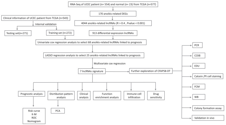
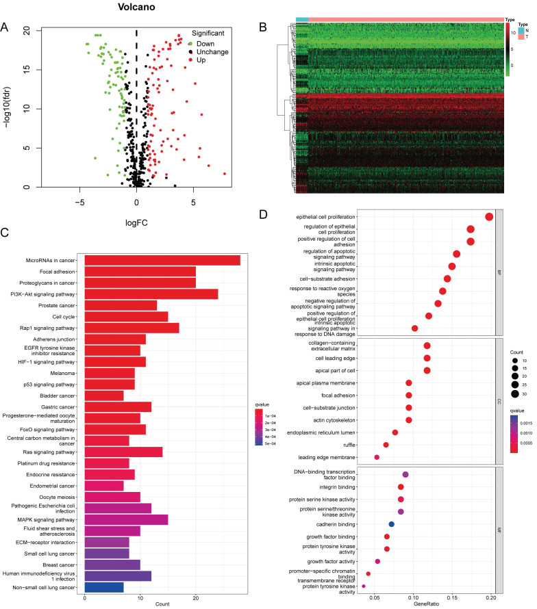
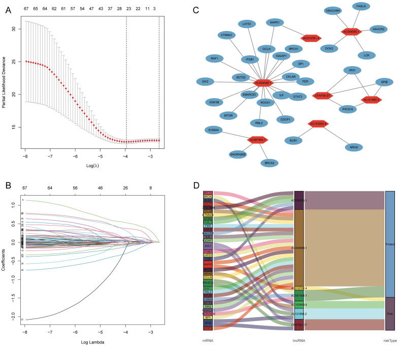
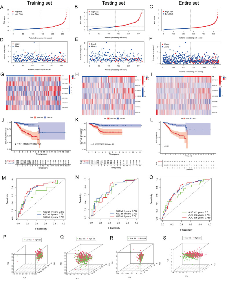
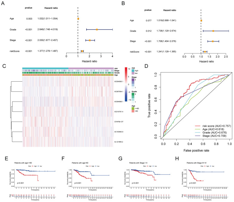
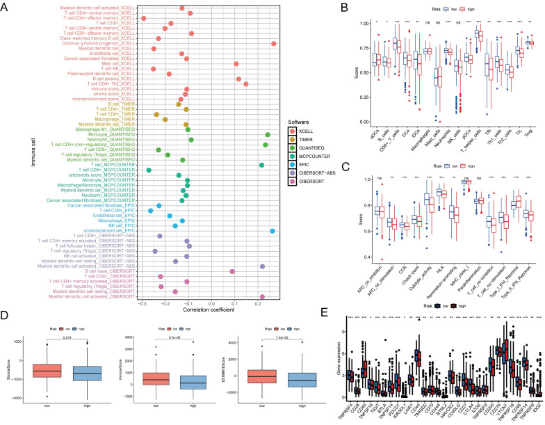
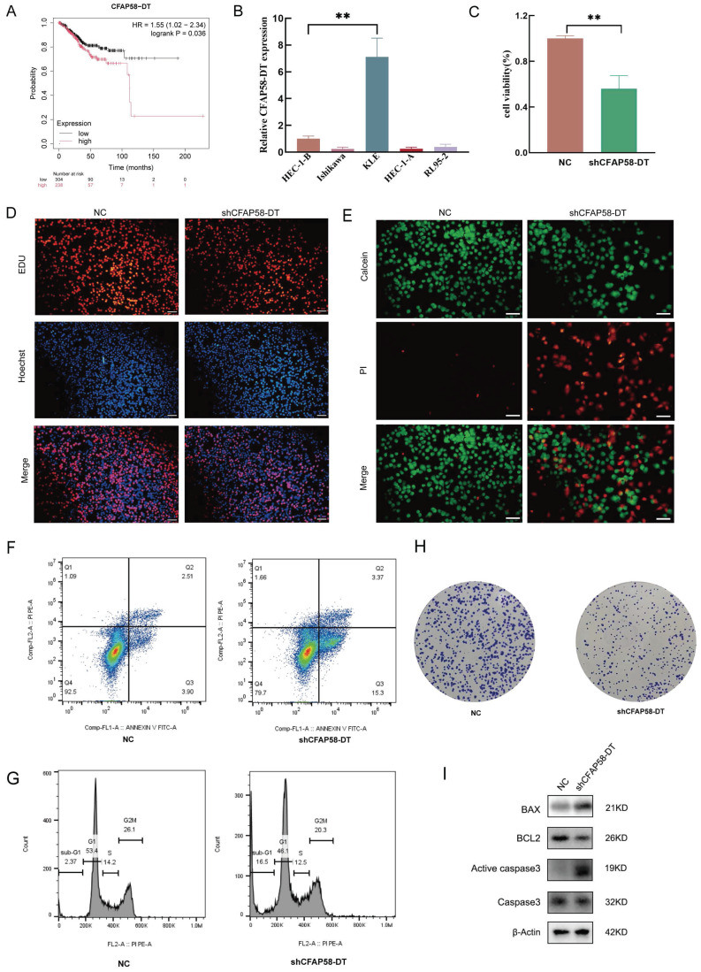
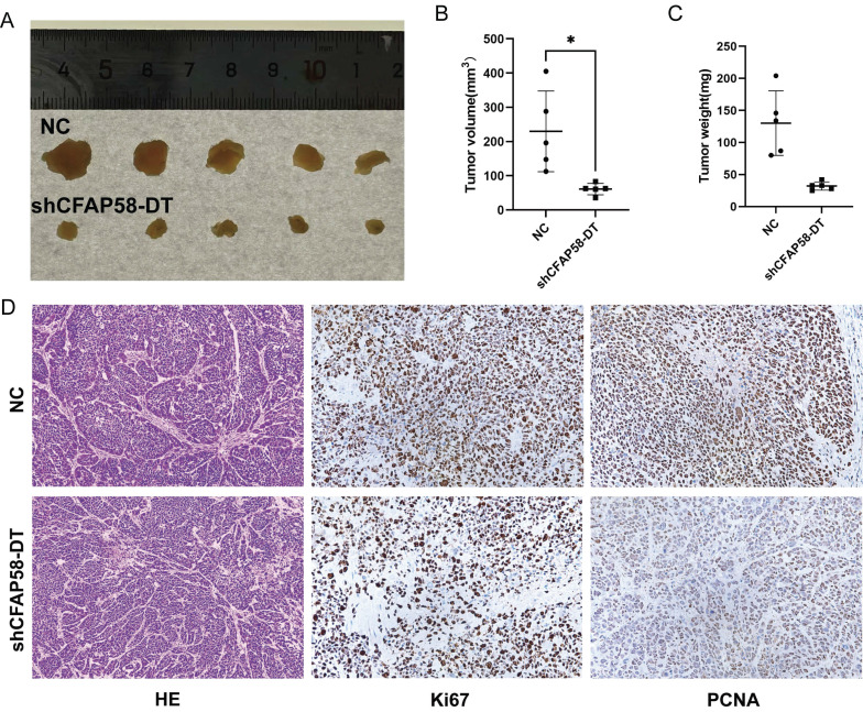
Background: Endometrial cancer is a highly heterogeneous malignancy in women with high mortality, and patients diagnosed with advanced endometrial cancer have a poor prognosis. Anoikis is a form of programmed cell death that is important for cancer development and metastasis. Long non-coding RNAs (lncRNAs) are considered critical regulators of gene expression and key players in cancer biology; however, the effects of anoikis-associated lncRNAs on the prognosis and treatment of patients with endometrial cancer remain unclear. Methods: Using transcriptome sequencing data and clinical information from The Cancer Genome Atlas database, we developed a novel prognostic signature for endometrial cancer based on anoikis-related lncRNAs by combining multivariate regression analysis and least absolute shrinkage and selection operator regression. The signature was validated by receiver operating characteristic (ROC) curve and Kaplan-Meier analyses. After analyzing the relationships between the seven lncRNAs in the signature and tumor progression through gene set enrichment analysis (GSEA), we further explored the differences in immune function and drug sensitivity. Additionally, to investigate the functions of these lncRNAs in the occurrence and development of endometrial cancer, we selected CFAP58-DT to conduct a series of in vitro and in vivo experiments to verify its partial functions. Results: Seven anoikis-associated lncRNAs (CFAP58-DT, AC004585.1, AC103563.9, AL121895.2, AC004596.1, AC010761.4, and AC087564.1) with prognostic value were identified for signature construction. The analysis showed excellent predictive accuracy of the signature (the largest area under the ROC curve = 0.757). GSEA indicated that these lncRNAs may regulate diverse cellular processes, including intercellular interactions, cell proliferation, differentiation, apoptosis, angiogenesis, glucose and fatty acid metabolism, immune responses, and inflammatory regulation. Furthermore, immune analysis revealed a high likelihood of immune evasion and poor immunotherapy efficacy in high-risk patients. However, there were distinct differences in the immune checkpoints and anticancer drug sensitivity between the two patient groups, which may aid in guiding treatment. Finally, our experiments showed that silencing CFAP58-DT significantly affected cell proliferation, promoted apoptosis, and reduced tumor malignancy. Conclusion: Our study highlights the significance of anoikis-associated lncRNAs in endometrial cancer progression and their potential as prognostic markers and therapeutic targets. The signature constructed using these lncRNAs may offer new avenues for endometrial cancer treatment and immunotherapy. The function of CFAP58-DT has been validated in vitro and in vivo, consistent with our previous analysis; however, further research into its upstream and downstream mechanisms is warranted.
Keywords: anoikis; endometrial cancer; lncRNAs; prognosis.
© The author(s).
Conflict of interest statement
Competing Interests: The authors have declared that no competing interest exists.
Figures
Similar articles
-
Identification of a PANoptosis-related long noncoding rna risk signature for prognosis and immunology in colon adenocarcinoma.BMC Cancer. 2025 Apr 10;25(1):662. doi: 10.1186/s12885-025-14021-2. BMC Cancer. 2025. PMID: 40211224 Free PMC article.
-
The disulfidptosis-related lncRNAs can predict survival and immunotherapy response accurately in endometrial carcinoma.Cell Mol Biol (Noisy-le-grand). 2025 Apr 15;71(3):20-30. doi: 10.14715/cmb/2025.71.3.3. Cell Mol Biol (Noisy-le-grand). 2025. PMID: 40235334
-
Identification and validation of a seven m6A-related lncRNAs signature predicting prognosis of ovarian cancer.BMC Cancer. 2022 Jun 8;22(1):633. doi: 10.1186/s12885-022-09591-4. BMC Cancer. 2022. PMID: 35676619 Free PMC article.
-
A Comprehensive Review of Long Non-Coding RNAs in the Cancer-Immunity Cycle: Mechanisms and Therapeutic Implications.Int J Mol Sci. 2025 May 17;26(10):4821. doi: 10.3390/ijms26104821. Int J Mol Sci. 2025. PMID: 40429961 Free PMC article. Review.
-
Association of long non-coding RNA in lipid metabolism: Implications in leukemia.Int J Biochem Cell Biol. 2025 Jul;184:106785. doi: 10.1016/j.biocel.2025.106785. Epub 2025 Apr 15. Int J Biochem Cell Biol. 2025. PMID: 40246061 Review.
References
-
- Crosbie EJ, Kitson SJ, McAlpine JN, Mukhopadhyay A, Powell ME, Singh N. Endometrial cancer. Lancet. 2022;399:1412–28. - PubMed
-
- Siegel RL, Miller KD, Fuchs HE, Jemal A. Cancer statistics, 2022. CA Cancer J Clin. 2022;72:7–33. - PubMed
-
- Tung HJ, Huang HJ, Lai CH. Adjuvant and post-surgical treatment in endometrial cancer. Best Pract Res Clin Obstet Gynaecol. 2022;78:52–63. - PubMed
MeSH terms
Substances
LinkOut - more resources
Full Text Sources
