[Preprint]. 2025 May 5:rs.3.rs-5849564.
doi: 10.21203/rs.3.rs-5849564/v2.

RPE65 variant p.(E519K) causes a novel dominant adult-onset maculopathy in 83 affected individuals

Affiliations

RPE65 variant p.(E519K) causes a novel dominant adult-onset maculopathy in 83 affected individuals

Eline Van Vooren et al. Res Sq. .

Abstract

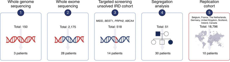
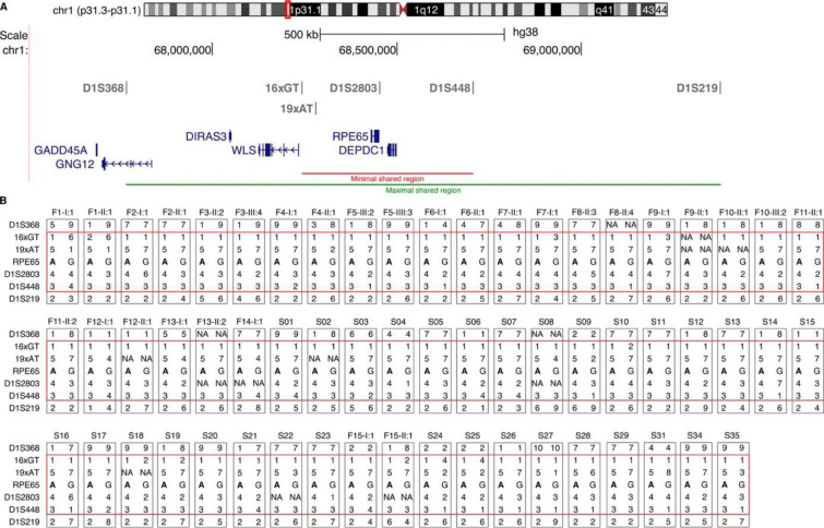
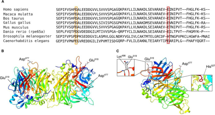
Recessive RPE65-related retinopathy is an inherited retinal disease (IRD) that is a well-established target for gene therapy. Dominant RPE65-related retinopathy, however, due to Irish founder variant p.(D477G), is extremely rare. Here, we report the discovery, replication and characterization of a novel dominant retinopathy caused by RPE65 variant p.(E519K), identified in 83 individuals of European ancestry across IRD registries (Belgian discovery cohort, n=2,873; replication cohort, n=18,796). Long-read sequencing-based haplotyping revealed a shared region of 464 kb, supporting a founder effect. Genotype-phenotype data support dominant inheritance and phenotypic variability respectively, characterized by late-onset macular dystrophy with two main subtypes, a pathognomonic mottled subtype and a pattern dystrophy subtype. Functional studies showed that the p.(E519K) variant affects RPE65 enzymatic activity, correlating with lower protein expression. Protein modelling and cellular thermal shift assays further supported a destabilizing effect on protein structure. Overall, our work provides strong genetic, clinical, molecular and functional evidence for a novel dominant RPE65 retinopathy in multiple families in Europe and North America due to a Belgian founder variant. This discovery reduces the diagnostic gap in dominant IRD, particularly in individuals of European ancestry. Finally, it lays the foundation for developing therapeutic strategies targeting dominant RPE65 retinopathy.

Keywords: Autosomal dominant; European ancestry; Founder; Inherited Retinal Disease (IRD); RPE65.


Conflict of interest statement

Conflicts of Interests The authors declare no competing interests

Figures

Figure 1.
Figure 1.. Workflow through which monoallelic patients for RPE65 c.1555G>A p.(E519K) were identified.
(1–4) represent the Belgian IRD discovery cohort: WGS analysis (n=150) and a second step of WES analysis (n=2,175) in IRD cases revealed c.1555G>A, p.(E519K) in 3 and 28 cases, respectively. Targeted testing of IRD patient cohorts that tested negative for mitochondrial retinopathies (MIDD), ABCA4-, BEST1-, and PRPH2-associated maculopathy (n=518) identified 14 more patients. Segregation analysis (n=51) showed segregation of p.(E519K) with disease in 30 affected family members. The total number of monoallelic p.(E519K) IRD cases in Belgian discovery cohort: n=75. (5) represents the IRD replication cohort (n=18,796). p.(E519K) was mined in IRD registries from France, The Netherlands, Germany, United Kingdom (UK), Scotland, Ireland, Canada. The total number of monoallelic p.(E519K) IRD cases found in the replication cohort: n=8, specifically France (n=3; of which one included in this study), the Netherlands (n=1), Canada (n=6). Overall number of monoallelic p.(E519K) IRD cases: n=85, of which 83 are included in this study.
Figure 2.
Figure 2.. Haplotype reconstruction in all Belgian index patients, one family member (when available) and the French (S34), Dutch (S35) and Canadian (S31) index cases.
(A) The location of the six microsatellite markers is depicted above a schematic representation of the genes in this region (chr1: 67611911–69469522). Red line: minimal shared region (464 kb), green line: maximal shared region (1.6 Mb). (B) Results from haplotype analysis. Each grey box represents the two alleles with the patient identifier shown at the top. The c.1555G>A (p.(E519K) variant is indicated in bold and the common allele is shown on the left. The red box highlights the shared markers. NA values: missing data. The conversion key for the microsatellite alleles can be found in Supplementary Table 9.
Figure 3.
Figure 3.. Conservation and protein modelling using crystal structures of RPE65.
(A) The final segment of the amino acid alignment for RPE65 of six vertebrate species and the related carotenoid cleavage dioxygenases from two invertebrate species is displayed. Orange box: Asp477; red box: Glu519. (B) In the 3FSN crystal structure, RPE65 is observed as a dimer. Both Asp477 and Glu519 are distant from the dimerization site. (C) The monomer crystal structure of RPE65 (4RYX), whereby a positively charged lysine (Lys498) is positioned in the vicinity of Glu519. If the glutamate is replaced by a lysine these residues (Lys498 and Lys519) will repel each other due to their positive charges. His527, important for iron binding in the catalytic center, could be affected by p.(E519K).
Figure 4.
Figure 4.. Variant p.(E519K) affects expression, enzymatic activity and thermal stability of RPE65.
(A) RPE65 protein expression following single or co-transfection of vectors encoding WT-RPE65, p.(D477G), or p.(E519K) proteins in HEK293-T cells. A shift was observed for the p.(D477G) variant. CRALBP and ß-tubulin protein expression were used as transfection and loading controls. NTC: no template control. (B) Relative RPE65 protein expression, normalized using CRALBP protein expression, is shown. A significant decrease in protein expression is observed after overexpression of p.(D477G) and p.(E519K), compared to WT-RPE65. Data are represented as median ± IQR. n = 18 for WT, p.(D477G) and p.(E519K), n=9 for WT+p.(D477G) and WT+p.(E519K), * is p<0.05 according to a Kruskal-Wallis test with Dunn’s multiple comparisons test. (C) p.(E519K) RPE65 showed reduced retinol isomerase activity (56%) compared to WT-RPE65 when measured in an in cellulo minimal visual cycle assay. (D) In co-IP experiments, ARPE-19 cells were either single or co-transfected with constructs encoding HA-tagged WT-RPE65 and/or MYC-tagged p.(D477G) or p.(E519K) protein. Co-IP was performed, using the anti-HA and anti-MYC antibodies and western blotting with either anti-HA or anti-MYC antibody was used to determine coprecipitation. The results demonstrate an interaction between the D477 and WT-RPE65 proteins, but not between p.(E519K) and WT-RPE65 proteins. Input represents total protein extract, FT (flow-through) the leftover unbound fraction, and B (bound) proteins specifically bound by the antibodies and precipitated by the beads. (E-F) Results from a CETSA assay to assess thermal stability after transfection of WT-RPE65 (E) and co-transfection of WT and p.(E519K) (F) in HEK293-T cells. The IC50, the temperature at which 50% of the protein is denatured and aggregated, was lowered in the WT + p.(E519K) condition. Immunoblots are depicted above the graphs, labels 1–12 represent the temperatures at which the cells were incubated (30, 37, 39.8, 42.8, 46.3, 48.8, 50.8, 54.1, 58.7, 62.1, 65.0 and 66.9°C, respectively). A non-linear regression was performed on the data and resulting means with standard deviation are shown in the graphs. R2 quantifies goodness of fit.
Figure 5.
Figure 5.. SW-AF imaging (top) and color fundus imaging (bottom) illustrating the representative distinct p.(E519K)-associated phenotypes: mottled phenotype (A-E) and pattern dystrophy (F-J).
(A) Left eye of 31-year-old female (F12-II:1). (B) Right eye of 41-year-old male (S02). (C) Right eye of 43-year-old male (F11-II:2). (D) Left eye of 29-year-old female (F2-III:1). (E) Right eye of 47-year-old female (F11-II:1). (F) Right eye of 59-year-old female (S10). (G) Right eye of 78-year-old male (F4-I:1). (H) Right eye of 72-year-old female (F3-II:2). (I) Left eye of 62-year-old female (F5-III:7). (J) Left eye of 58-year-old male (F8-II:5). SW-AF = short-wavelength autofluorescence.

References

    1. Bourne RRA, Steinmetz JD, Saylan M, et al. Causes of blindness and vision impairment in 2020 and trends over 30 years, and prevalence of avoidable blindness in relation to VISION 2020: The Right to Sight: An analysis for the Global Burden of Disease Study. Lancet Glob Health. 2021;9(2):e144–e160. doi:10.1016/S2214-109X(20)30489-7 - DOI - PMC - PubMed
    1. WHO. Blindness and vision impairment. August 10, 2023. Accessed October 1, 2023. https://www.who.int/news-room/fact-sheets/detail/blindness-and-visual-im...
    1. Redmond TM, Yu S, Lee E, et al. Rpe65 is necessary for production of 11-cis-vitamin A in the retinal visual cycle. Nat Genet. 1998;20(4):344–351. doi:10.1038/3813 - DOI - PubMed
    1. Redmond TM, Poliakov E, Yu S, Tsai JY, Lu Z, Gentleman S. Mutation of key residues of RPE65 abolishes its enzymatic role as isomerohydrolase in the visual cycle. Proc Natl Acad Sci U S A. 2005;102(38):13658–13663. doi:10.1073/pnas.0504167102 - DOI - PMC - PubMed
    1. Moiseyev G, Chen Y, Takahashi Y, Wu BX, Ma JX. RPE65 Is the Isomerohydrolase in the Retinoid Visual Cycle.; 2005. www.pnas.orgcgidoi10.1073pnas.0503460102 - PMC - PubMed

Publication types

LinkOut - more resources