. 2025 Feb 24;4(3):lnaf007.
doi: 10.1093/lifemedi/lnaf007. eCollection 2025 Jun.

Follicular development of fetal gonads under the skin of adult mice

Affiliations

Follicular development of fetal gonads under the skin of adult mice

Jiyu Chen et al. Life Med. .

Abstract

Adult ovarian tissues or biopsies isolated from patients prior to chemotherapy or irradiation can reconstitute ovarian functions when transplanted either in the abdomen or subcutaneously. Subcutaneously transplantation avoids invasive surgery and potential risks associated with internal procedures. We investigated whether functional ovaries could develop subcutaneously from early E12.5 fetal gonads without entering meiosis in mouse model. Unexpectedly, the subcutaneously transplanted fetal gonads failed to undergo folliculogenesis in the recipient mice. The transplanted gonads experienced meiotic deficiency and exhibited significant defects in DNA repair and recombination, increased apoptosis levels. Meiotic defects in the subcutaneous grafts were partly attributable to variations in temperature and oxygen concentration. However, completion of meiotic prophase I was effectively achieved through in vitro culture of the gonads at 37°C. Subsequently, the in vitro cultured E12.5 gonads, following subcutaneous transplantation, became competent in folliculogenesis, restoring endocrine functions. This finding may have implications for rejuvenating ovarioids from fetal gonad-like cells using pluripotent stem cell technologies, as well as for enhancing endocrine recovery and health span.

Keywords: follicle development; gonad; meiosis; ovary; subcutaneous transplantation.


Conflict of interest statement

The authors declare no competing interests.

Figures

Figure 1.
Figure 1.
Inability of E12.5 gonads to form follicular structures following subcutaneous transplantation.(A) Morphology of a 3-week-old ovary, a gonad cultured in vitro for 25 days (in vitro), a 25-day transplantation in KC, and a 25-day graft transplanted beneath the skin (SC). Scale bar: 200 μm. (B) Area measurements of the 3-week-old ovary, 25-day in vitro cultured gonad, KC graft and SC graft. n ≥ 4. Mean ± SD. (C) Immunofluorescence staining of Ddx4 (green) and Foxl2 (red) in 3-week-old ovaries, 25-day in vitro cultured gonads, KC grafts and SC grafts. Scale bar: 100 µm. (D) Three-week-old ovaries, in vitro cultured gonads, KC grafts and SC grafts were embedded in paraffin, and 8-μm-thick sections were prepared and stained with H&E. Scale bar: 200 µm. (E) The numbers of follicles per 3-week-old ovary, in vitro cultured gonad, KC grafts and SC grafts were counted. Combining the counts from every fifth section over the entire tissue yielded the total number of follicles in the tissue. n ≥ 3. Mean ± SD. (F) Percentage of different types of follicles per sample. n ≥ 3. Mean ± SD. *P < 0.05, **P < 0.01, ***P < 0.001, ns = not significant. Tukey’s multiple comparisons test for (B), (E), (F).
Figure 2.
Figure 2.
Meiotic defects in subcutaneous grafts.(A) Immunofluorescence staining of Stra8 (red) of the 2- and 4-day in vitro cultured gonad, KC graft and SC graft transplanted for two and four days. Scale bar: 20 µm. The number of Stra8-positive cells in in vitro cultured gonads, KC grafts and SC grafts was counted. n ≥ 3. Mean ± SD. (B) Meiocyte spread and immunofluorescence staining of SYCP1 (green) and SYCP3 (red) in E16.5 female gonads, in vitro cultured gonads, KC transplants and SC transplants at 4 days. Scale bar: 20 μm. (C) Percentage of SYCP3+ cells in E16.5 female gonads, in vitro cultured gonads, a 4-day KC transplant and SC transplant. Mean ± SD. (D) Percentage of normal number of synaptonemal complexes at pachytene in gonad cultured in vitro, a 4-day KC graft and SC graft. Mean ± SD. (E) Percentage of different developmental stages of meiotic cells in cultured gonads and grafts. *P < 0.05, **P < 0.01, ***P < 0.001, ns = not significant. ANOVA for (A) and Tukey’s multiple comparisons test for (C),(D), (E).
Figure 3.
Figure 3.
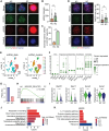
Meiocytes in SC grafts with DSB repair defects during meiotic prophase I.(A) Representative images of immunofluorescence staining of SYCP3 (green) and γH2AX (red) in pachytene stage meiocytes from KC transplants and SC transplants, respectively. Representative image of meiocytes with severe γH2AX signals in SC transplants. Scale bar: 10 μm. (B) Fluorescence intensity of γH2AX in KC transplants and SC transplants. Quantification of the fluorescence intensity in pachytene cells was performed using ImageJ under the same parameters. Data are presented as mean ± SD. (C) Percentage of meiocytes with severe γH2AX intensity in γH2AX-positive cells in KC and SC transplants. Mean ± SD. (D) Representative images of immunofluorescence staining of SYCP3 (red) and RAD51 (green) in pachytene stage meiocytes from KC transplants and SC transplants. Scale bar: 10 μm. Quantification of RAD51 foci number in pachytene stage meiocytes from KC transplants and SC transplants. (E) Two-dimensional (2D) visualization of clusters based on different samples (left) and transcriptional patterns (right) using UMAP. (F) Dot plot of the PGC, pregranulosa cell, mesothelial cell, endothelial cell and interstitial cell marker expression across all cell types. Each cell cluster is color-coded by identity. The dot size represents the percentage of cells expressing the indicated genes in each cluster, while dot color intensity reflects the average expression level of the indicated genes. (G) Percentage of PGC across different samples. (H) GSEA of meiosis-related pathways. (I) The VlnPlot showing differentially expressed genes associated with meiosis. (J) Top enriched GO terms of DEGs between KC and SC in PGC. (K) Top enriched KEGG terms of DEGs between KC and SC in PGC. Mean ± SD. *P < 0.05, **P < 0.01, ***P < 0.001, ns = not significant. Student’s t test for (B), (C), (D).
Figure 4.
Figure 4.
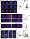
The effect of temperature and oxygen concentration on meiotic prophase I in SC grafts.(A) Meiocyte spreading and immunofluorescence staining of SYCP1 (green) and SYCP3 (red) in gonads cultured at 37°C, gonads cultured at 33°C and six-day SC transplants. Scale bar: 10 μm. (B) Percentage of SYCP3-positive cells in SC-transplanted gonads at 6 days, compared to gonads cultured at 37°C and 33°C. The proportion of SYCP3+ cells to total cells was calculated in each field of view. Mean ± SD. (C) Meiotic cells at various developmental stages in SC grafts and gonads cultured at 37°C and 33°C, respectively. Mean ± SD. (D) Immunofluorescence staining of Ddx4 in gonads co-cultured with fat at 33°C for 4 and 6 days. Scale bar: 20 µm. The number of Ddx4-positive cells was counted. Mean ± SD. (E) Immunofluorescence staining of Stra8 in gonads co-cultured with fat at 33°C for 4 days. Scale bar: 20 µm. The number of Stra8-positive cells was counted. Mean ± SD. (F) Meiocyte spreading and immunofluorescence staining of SYCP1 and SYCP3 in gonads cultured in 20% oxygen and 5% oxygen, respectively. Scale bar: 20 µm. (G) Percentage of SYCP3-positive cells in SC-transplanted gonads at 6 days and in gonads cultured in 20% oxygen and 5% oxygen. (H) Meiotic cells at various developmental stages in SC grafts and in gonads cultured in 20% oxygen and 5% oxygen, respectively. Mean ± SD. *P < 0.05, **P < 0.01, ***P < 0.001, ns = not significant. Tukey’s multiple comparisons test for (B), (C),(G), (H). Student’s t test for (D),(E).
Figure 5.
Figure 5.
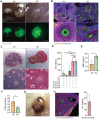
Folliculogenesis of short-term cultured fetal gonads following SC transplantation.(A) Morphology of 25-day grafts at both the KC and SC sites after the transplantation of an 8-day in vitro cultured gonad. Scale bar: 1 mm. (B) Immunofluorescence staining of Ddx4 and Foxl2 in 25-day grafts at both KC and SC sites following the transplantation of an 8-day in vitro cultured gonad. Scale bar: 20 µm. (C) H&E staining of a 25-day graft at both the KC and SC sites after transplantation of an 8-day in vitro cultured gonad. Scale bar: 100 µm. (D) Follicle count in 25-day grafts at the SC site after the transplantation of 2-, 4-, 6- and 8-day in vitro cultured E12.5 gonad. n ≥ 3. Mean ± SD. (E) Serum AMH levels in mice with SC grafts compared to mice with bilateral OE. n = 3. Mean ± SD. (F) Serum FSH levels in mice with subcutaneous grafts compared to mice with bilateral OE. n = 3. Mean ± SD. (G) Morphology of an 8-week graft at the SC site following the transplantation of an 8-day in vitro cultured gonad. Scale bar: 1 mm. (H) Immunofluorescence staining of Ddx4 and Foxl2 in 8-week grafts at the SC site after the transplantation of an 8-day in vitro cultured gonad. Scale bar: 20 µm. The number of follicles in 8-week gonads at the SC site transplanted under the skin for 8 weeks. n ≥ 3. Mean ± SD. *P < 0.05, **P < 0.01, ***P < 0.001, ns = not significant. Tukey’s multiple comparisons test for (D). Student’s t test for (E), (F).
Figure 6.
Figure 6.
Follicular development of fetal gonads under the skin of adult mice.Follicular development in fetal gonads under adult mouse skin is hindered by meiotic defects due to unsuitable temperature and oxygen levels. However, short-term in vitro culture of E12.5 female gonads in optimal conditions allows them to complete meiosis, enabling functional egg formation after subcutaneous transplantation.

Similar articles

References

    1. Christensen K, Doblhammer G, Rau R, et al.Ageing populations: the challenges ahead. Lancet 2009;374:1196–208. - PMC - PubMed
    1. Inter LST. Variations in reproductive events across life: a pooled analysis of data from 505 147 women across 10 countries. Hum Reprod 2019;34:881–93. - PMC - PubMed
    1. Xiong J, Kang SS, Wang Z, et al.FSH blockade improves cognition in mice with Alzheimer’s disease. Nature 2022;603:470–6. - PMC - PubMed
    1. Zhu Z, Xu W, Liu L.. Ovarian aging: mechanisms and intervention strategies. Medical review (2021) 2022;2:590–610. - PMC - PubMed
    1. Lobo RA. Hormone-replacement therapy: current thinking. Nat Rev Endocrinol 2017;13:220–31. - PubMed

LinkOut - more resources