

C3HeB/FeJ mice with chronic Mycobacterium avium complex pulmonary infection exhibit impaired respiratory function but not necrotising granulomatous disease
- PMID: 40386578
- PMCID: PMC12081485
- DOI: 10.1186/s44350-025-00004-7
C3HeB/FeJ mice with chronic Mycobacterium avium complex pulmonary infection exhibit impaired respiratory function but not necrotising granulomatous disease
Abstract
Background: Mycobacterium avium complex (MAC) is driving a global rise in pulmonary disease (MAC-PD) characterised by chronic infection, granulomatous inflammation and impaired respiratory function. Better animal models are needed to screen candidate therapies targeting bacteria and immune-mediated tissue injury. The C3HeB/FeJ mouse was previously reported to model necrotic granulomatous lung infection in MAC-PD following infection with a low-dose inoculum of the clinical isolate MAC2285R. We investigated whether this model was reproducible with variations in MAC strain and inoculating dose.
Methods: Six-week-old female C3HeB/FeJ mice were infected intratracheally with a clinical isolate of MAC (MAC2285R) or reference strains (MAC104 or MAC101). Mice were culled at 4-weekly intervals post-infection until week 12. Lungs, spleen and liver were harvested for bacterial burden enumeration and histological examination. Whole body plethysmography (WBP) was performed weekly to measure changes in respiratory function (Buxco system).
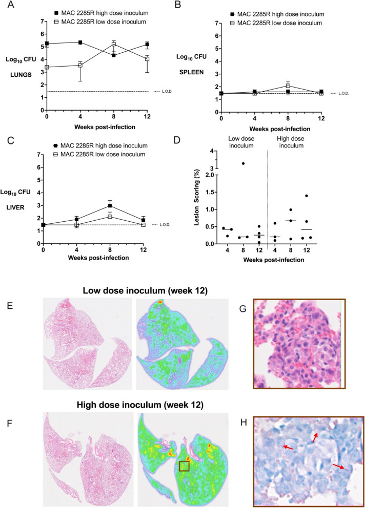
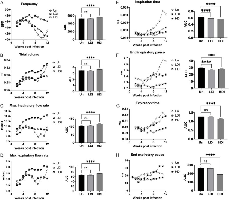
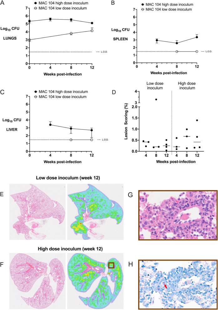
Results: C3HeB/FeJ mice infected with low dose inoculum of MAC2285R infection exhibited increasing bacterial lung infection for 8 weeks (p < 0.05), followed by stable lung burden from weeks 8-12. High dose inoculum resulted in stable lung bacterial burden over 12 weeks. Histological analysis revealed only mild inflammatory changes in both low and high dose inoculum groups at weeks 4, 8 and 12 post-infection, with no evidence of necrotising or non-necrotising granulomatous inflammation. Surrogate measures of respiratory effort (frequency, tidal volume, inspiratory and expiratory flow rates) were increased in mice with high dose inoculum compared to uninfected controls (p < 0.001), but not low dose inoculum. Similar findings on lung bacterial burden and histological analysis were found in mice infected with low- and high-dose inoculum of MAC104 and MAC101. MAC104 infection caused greater changes in respiratory function, whereas MAC101 did not significantly affect breathing patterns.
Conclusion: The C3HeB/FeJ mouse is susceptible to chronic MAC infection from intratracheal infection with reference and clinical isolates, but this was not associated with severe granulomatous inflammation as previously reported. A low dose inoculum generated a proliferative lung infection, whereas high dose inoculum resulted in chronic, stable lung bacterial burden. Mice with high-dose inoculum MAC2285R and MAC104 infection also displayed evidence of increased respiratory effort.
Supplementary information: The online version contains supplementary material available at 10.1186/s44350-025-00004-7.
Keywords: Animal model; Granuloma; Mycobacterium avium complex; Plethysmography; Respiratory infection.
© The Author(s) 2025.
Conflict of interest statement
Competing interests The authors declare no competing interests.
Figures


Similar articles
-
Mycobacterium avium Infection in a C3HeB/FeJ Mouse Model.Front Microbiol. 2019 Apr 3;10:693. doi: 10.3389/fmicb.2019.00693. eCollection 2019. Front Microbiol. 2019. PMID: 31001241 Free PMC article.
-
Pulmonary granuloma formation during latent Cryptococcus neoformans infection in C3HeB/FeJ mice involves progression through three immunological phases.mBio. 2025 Feb 5;16(2):e0361024. doi: 10.1128/mbio.03610-24. Epub 2025 Jan 14. mBio. 2025. PMID: 39807873 Free PMC article.
-
Novel Screening System of Virulent Strains for the Establishment of a Mycobacterium avium Complex Lung Disease Mouse Model Using Whole-Genome Sequencing.Microbiol Spectr. 2022 Jun 29;10(3):e0045122. doi: 10.1128/spectrum.00451-22. Epub 2022 May 17. Microbiol Spectr. 2022. PMID: 35579455 Free PMC article.
-
[Clinical study on development of nontuberculous mycobacterial lung disease].Kekkaku. 2004 Dec;79(12):737-41. Kekkaku. 2004. PMID: 15782619 Review. Japanese.
-
[Mycobacteriosis as zoonotic disease--comparative pathological study on Mycobacterium avium complex infection].Kekkaku. 2007 Jun;82(6):539-50. Kekkaku. 2007. PMID: 17633122 Review. Japanese.
References
-
- Kwak N, Park J, Kim E, Lee CH, Han SK, Yim JJ. Treatment Outcomes of Mycobacterium avium Complex Lung Disease: A Systematic Review and Meta-analysis. Clin Infect Dis. 2017;65:1077–84. - PubMed
-
- Mehta M, Chapman KR, Heffer M, Marras TK. Impact of pulmonary nontuberculous mycobacterial treatment on pulmonary function tests in patients with and without established obstructive lung disease. Respirology. 2015;20:987–93. - PubMed
-
- Fujita J, Ohtsuki Y, Shigeto E, Suemitsu I, Yamadori I, Bandoh S, et al. Pathological findings of bronchiectases caused by Mycobacterium avium intracellulare complex. Respir Med. 2003;97:933–8. - PubMed
LinkOut - more resources
Full Text Sources