. 2025 Apr 30;16(2):679-698.
doi: 10.21037/jgo-24-685. Epub 2025 Apr 27.

Downregulation of CPT2 promotes proliferation and migration through the TNFα/NF-κB pathway in cholangiocarcinoma

Affiliations

Downregulation of CPT2 promotes proliferation and migration through the TNFα/NF-κB pathway in cholangiocarcinoma

Jun Mao et al. J Gastrointest Oncol. .

Abstract

Background: Carnitine palmitoyltransferase II (CPT2) is an important regulatory enzyme involved in fatty acid oxidation; it is associated with the prognosis and progression of colorectal and ovarian cancers, but its expression and role in cholangiocarcinoma (CCA) have been less explored. This study aims to explore the role and molecular mechanism of CPT2 in CCA and to determine the potential relationship between CPT2 expression and the prognosis of CCA patients.

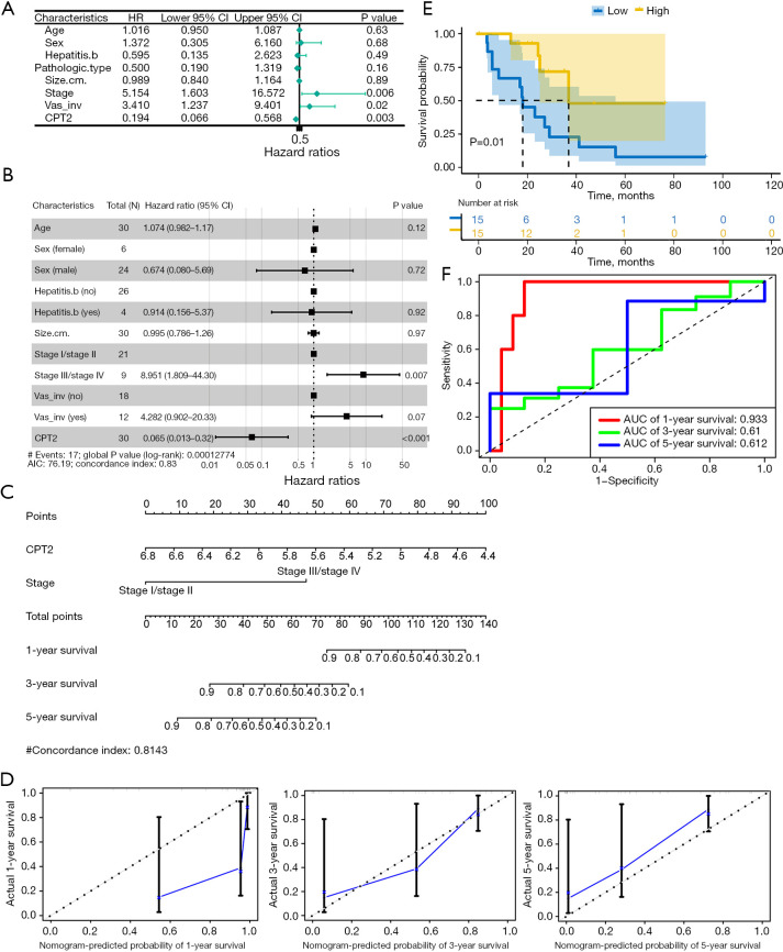
Methods: Bioinformatics analyses were used to assess CPT2 expression in CCA and other cancers. Independent prognostic factors of CCA were identified for univariate and multivariate Cox regression analyses. Nomograms were employed to predict CCA 1-, 3-, and 5-year survival. Kaplan-Meier curves explored the correlation between CPT2 expression and CCA survival. We used time-dependent receiver operating characteristics (ROCs) to evaluate the predictive efficiency of CPT2. Furthermore, potential mechanisms of CPT2 were analyzed by Gene Set Enrichment Analysis (GSEA) in CCA. CPT2 expression in peripheral blood, tissues, and cell lines of CCA was verified by quantitative real-time polymerase chain reaction (qRT-PCR) and Western blotting. The effect of CPT2 on CCA cells was gauged using Cell Counting Kit-8 (CCK-8), cell cycle, apoptosis, and transwell assays. Finally, the regulation of the TNFα/NF-κB pathway by CPT2 was verified by Western blotting.

Results: CPT2 expression was down-regulated in many cancers, including CCA. COX regression analyses showed that CPT2 expression and the clinical stage could be independent prognostic factors in CCA. Nomograms indicated that the lower probability of CCA survival was associated with the lower expression of CPT2 and the higher clinical stage. The Kaplan-Meier curve showed that the low expression of CPT2 was related to a poor prognosis in CCA. The time-dependent ROC curve demonstrated the predictive ability of CPT2 [1-, 3-, 5-year are under the curve (AUC) =0.933, 0.61, 0.612]. Functionally, CPT2 overexpression inhibited CCA cell proliferation, down-regulated CDK4/6 expression to arrest CCA cells at G1, induced apoptosis by up-regulating BAX expression, cleaved-caspase-3 expression, and down-regulating Bcl2 expression, and reduced migration and invasion via suppression of epithelial-mesenchymal transition (EMT). Knocking down CPT2 showed the opposite results. Mechanistically, overexpression of CPT2 could decrease TNFα and phosphorylated p65 (p-p65; Ser536) expression and inhibit NF-κB pathway activation. CPT2 knockdown yielded opposite results.

Conclusions: CPT2 is a potential prognostic marker of CCA, a tumor suppressor gene to inhibit the malignant progression of CCA, and therefore a potential therapeutic target.

Keywords: Cholangiocarcinoma (CCA); NF-κB; bioinformatics; carnitine palmitoyltransferase II (CPT2).


Conflict of interest statement

Conflicts of Interest: All authors have completed the ICMJE uniform disclosure form (available at https://jgo.amegroups.com/article/view/10.21037/jgo-24-685/coif). The authors have no conflicts of interest to declare.

Figures

Figure 1
Figure 1
CPT2 expression was down-regulated in CCA and other cancers. (A,B) The expression of CPT2 was significantly lower in CCA cancer tissues than in adjacent normal tissues in (A) GEO, and (B) TCGA datasets. (C) Pan-cancer analysis of CPT2. (D,E) The mRNA level of CPT2 was measured by qRT-PCR in (D), CCA peripheral blood and (E), 11 pairs of CCA tissues. (F) Protein expression of CPT2 was measured by Western blotting in 9 pairs of CCA tissues. The differences were analyzed with two-tailed Student t-tests or paired t-tests. The data are presented as the mean ± SD. *, P<0.05; **, P<0.01; ***, P<0.001; ****, P<0.0001. Experiments were repeated three times. CCA, cholangiocarcinoma; CPT2, carnitine palmitoyltransferase II; GAPDH, glyceraldehyde-3-phosphate dehydrogenase; GEO, Gene Expression Omnibus; mRNA, messenger RNA; qRT-PCR, quantitative real-time polymerase chain reaction; SD, standard deviation; TCGA, The Cancer Genome Atlas; TPM, transcripts per million.
Figure 2
Figure 2
CPT2 was an independent prognostic factor of CCA and had a positive correlation with patient prognosis. (A) Univariate COX and (B) multivariate COX regression analyses of CPT2 and clinicopathological features. (C) A nomogram diagram and (D) calibration curve were constructed by CPT2 expression and clinical stage. (E) The Kaplan Meier curve of high and low expression groups of CPT2. (F) A time-dependent ROC curve for CPT2 predicted the 1-, 3-, and 5-year survival of patients with CCA. The differences were analyzed using the Cox proportional hazards model and log-rank tests. Low, low CPT2 expression group; High, high CPT2 expression group; AUC, area under the curve; CCA, cholangiocarcinoma; CI, confidence interval; CPT2, carnitine palmitoyltransferase II; HR, hazard ratio; ROC, receiver operating characteristic; vas_inv, vascular invasion.
Figure 3
Figure 3
The molecular mechanism of CPT2 in CCA was predicted by GSEA. (A-C) The results of GSEA showed that (A) HALLMARK_TNFA_SIGNALING_VIA_NFKB, (B) HALLMARK_HYPOXIA, and (C) HALLMARK_EPITHELIAL_MESENCHYMAL_TRANSITION were significantly enriched in the low CPT2 expression group. (D-G) Analyses of correlations between the expression of CPT2 and (D) NFKBIA, (E) NFKBIB, (F) NFKB1_S536, and (G) CDH2 in the cBioPortal database. The differences were analyzed using the GSEA and Spearman correlation analysis. CCA, cholangiocarcinoma; CPT2, carnitine palmitoyltransferase II; ES, enrichment score; FDR, false discovery rate; GSEA, Gene Set Enrichment Analysis; mRNA, messenger RNA; NES, normalized enrichment score; RSEM, RNA-seq by expectation-maximization.
Figure 4
Figure 4
Overexpression of CPT2 inhibited the proliferation of CCA. (A) mRNA and protein levels of CPT2 in CCA cell lines and HIBEpiC were analyzed by qRT-PCR and Western blotting. (B) The fluorescence expression of RBE cells was detected using a fluorescence microscope. Scale bars: 100 μm. (C,D) The overexpression efficiency of RBE and HCCC9810 cells was evaluated by qRT-PCR and Western blotting. (E) Cell viabilities of RBE and HCCC9810 cells with CPT2 overexpression were analyzed by CCK-8. The differences were analyzed with two-tailed Student t-tests. The data are presented as the mean ± SD. *, P<0.05; **, P<0.01; ***, P<0.001; ****, P<0.0001. Experiments were repeated three times. CCA, cholangiocarcinoma; CCK8, Cell Counting Kit-8; CPT2, carnitine palmitoyltransferase II; GAPDH, glyceraldehyde-3-phosphate dehydrogenase; HIBEpiC, human normal bile duct epithelial cell; mRNA, messenger RNA; qRT-PCR, quantitative real-time polymerase chain reaction; SD, standard deviation.
Figure 5
Figure 5
Overexpression of CPT2 arrested CCA cells in the G1 phase and induced apoptosis. (A,B) Cell cycles of the overexpressed CPT2 group and vector group were analyzed by flow cytometry. (C,D) Cell apoptosis of the overexpressed CPT2 group and vector group was analyzed by flow cytometry. (E,F) The expressions of (E) CDK4, CDK6, (F) Bcl2, Bax, caspase 3, and cleaved-caspase 3 in the overexpressed CPT2 group and vector group were analyzed by Western blotting. The differences were analyzed with two-tailed Student t-tests. The data are presented as the mean ± SD. *, P<0.05; **, P<0.01; ***, P<0.001; ****, P<0.0001. Experiments were repeated three times. G1, G1 phase of the cell cycle; G2, G2 phase of the cell cycle; S, S phase of the cell cycle; CCA, cholangiocarcinoma; CPT2, carnitine palmitoyltransferase II; SD, standard deviation.
Figure 6
Figure 6
Overexpression of CPT2 inhibited migration and invasion of CCA cells through inhibiting EMT. (A,B) The invasion and migration abilities of the overexpressed CPT2 group and vector group were analyzed by transwell assays and visualized by crystal violet staining. Scale bars: 100 μm. (C) The expressions of EMT pathway-related proteins in the overexpressed CPT2 group and vector group were analyzed by Western blotting. The differences were analyzed with two-tailed Student t-tests. The data are presented as the mean ± SD. *, P<0.05; **, P<0.01; ****, P<0.0001. Experiments were repeated three times. CCA, cholangiocarcinoma; CPT2, carnitine palmitoyltransferase II; EMT, epithelial-mesenchymal transition; SD, standard deviation.
Figure 7
Figure 7
CPT2 knockdown could promote the proliferation, migration, and invasion of CCA cells. (A,B) CPT2 knockdown efficiency of HCCC9810 was evaluated by (A) qRT-PCR and (B) Western blotting. (C) Cell viabilities of the CPT2 knockdown group and Scramble group were analyzed by CCK-8. The (D) cell cycle and (E) apoptosis of HCCC9810 cells with CPT2 knockdown were analyzed by flow cytometry. (F,G) The expressions of (F) CDK4, CDK6, (G) Bcl2, Bax, caspase 3, and cleaved-caspase 3 in HCCC9810 cells with CPT2 knockdown were analyzed by Western blotting. (H) The migration and invasion of HCCC9810 cells with CPT2 knockdown were analyzed by transwell assay and visualized by crystal violet staining. Scale bars: 100 μm. (I) The expressions of EMT-related proteins in HCCC9810 cells with CPT2 knockdown were analyzed by Western blotting. The differences were analyzed with two-tailed Student t-tests. The data are presented as the mean ± SD. *, P<0.05; **, P<0.01; ***, P<0.001; ****, P<0.0001. Experiments were repeated three times. G1, G1 phase of the cell cycle; G2, G2 phase of the cell cycle; S, S phase of the cell cycle; CCA, cholangiocarcinoma; CPT2, carnitine palmitoyltransferase II; CCK8, Cell Counting Kit-8; EMT, epithelial-mesenchymal transition; GAPDH, glyceraldehyde-3-phosphate dehydrogenase; qRT-PCR, quantitative real-time polymerase chain reaction; SD, standard deviation.
Figure 8
Figure 8
The inhibitory effect of CPT2 on CCA cells may be related to inhibition of the TNFα/NF-κB signaling pathway. (A-C) Expression of related proteins of the TNFα/NF-κB signaling pathway in CCA cells with CPT2 (A,B) overexpression, or (C) knockdown. (D) The specific process of CPT2 regulating the TNFα/NF-κB signaling pathway (created with BioRender.com). The differences were analyzed with two-tailed Student t-tests. The data are presented as the mean ± SD. *, P<0.05; **, P<0.01; ***, P<0.001. Experiments were repeated three times. CCA, cholangiocarcinoma; CPT2, carnitine palmitoyltransferase II; GAPDH, glyceraldehyde-3-phosphate dehydrogenase.

Similar articles

References

    1. Dai YS, Hu HJ, Lv TR, et al. The influence of resection margin width in patients with intrahepatic cholangiocarcinoma: a meta-analysis. World J Surg Oncol 2023;21:16. 10.1186/s12957-023-02901-5 - DOI - PMC - PubMed
    1. Krasinskas AM. Cholangiocarcinoma. Surg Pathol Clin 2018;11:403-29. 10.1016/j.path.2018.02.005 - DOI - PubMed
    1. Stüben BO, Ahmadi S, Saner FH, et al. The significance of resection margins on R0 results in intrahepatic cholangiocarcinoma. Surg Oncol 2024;53:102058. 10.1016/j.suronc.2024.102058 - DOI - PubMed
    1. Gupta A, Sahai P, Prasad M, et al. Treatment Response and Survival with Chemotherapy for Unresectable, Nonmetastatic Cholangiocarcinoma. Euroasian J Hepatogastroenterol 2024;14:5-8. 10.5005/jp-journals-10018-1396 - DOI - PMC - PubMed
    1. Zhang Y, Esmail A, Mazzaferro V, et al. Newest Therapies for Cholangiocarcinoma: An Updated Overview of Approved Treatments with Transplant Oncology Vision. Cancers (Basel) 2022;14:5074. 10.3390/cancers14205074 - DOI - PMC - PubMed

LinkOut - more resources