. 2025 Apr 30;14(4):1351-1370.
doi: 10.21037/tlcr-2025-267. Epub 2025 Apr 27.

LNCAROD was stabilized through N6-methyladenosine methylation and exerted its anticancer effects in lung squamous cell carcinoma by inhibiting SIRT1 activity via CCAR2

Affiliations

LNCAROD was stabilized through N6-methyladenosine methylation and exerted its anticancer effects in lung squamous cell carcinoma by inhibiting SIRT1 activity via CCAR2

Qihang Yan et al. Transl Lung Cancer Res. .

Abstract

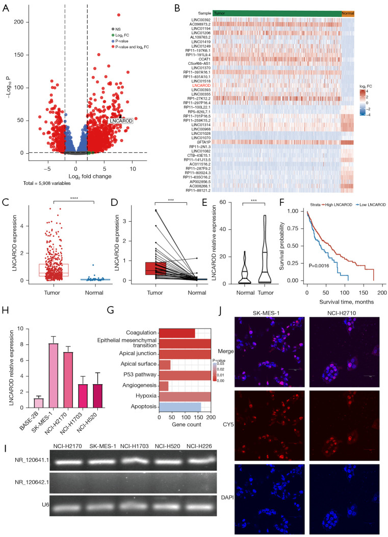
Background: Lung squamous cell carcinoma (LUSC), a deadly malignant tumor, is highly prevalent worldwide. Accumulating evidence indicates that long-chain noncoding RNAs play crucial regulatory roles in the occurrence and progression of LUSC. LNCAROD regulates the proliferation, migration, and invasion of cells by upregulating SERPINE1 expression in lung adenocarcinoma (LUAD). However, the functional mechanism of LNCAROD action in LUSC remains unclear. The aim of this study was to investigate the regulatory function and mechanism of LNCAROD action in the development of LUSC.

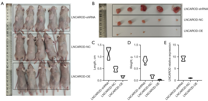
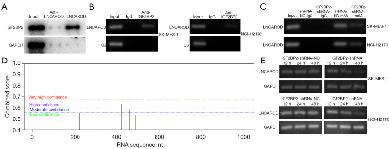
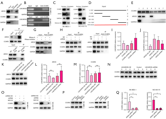
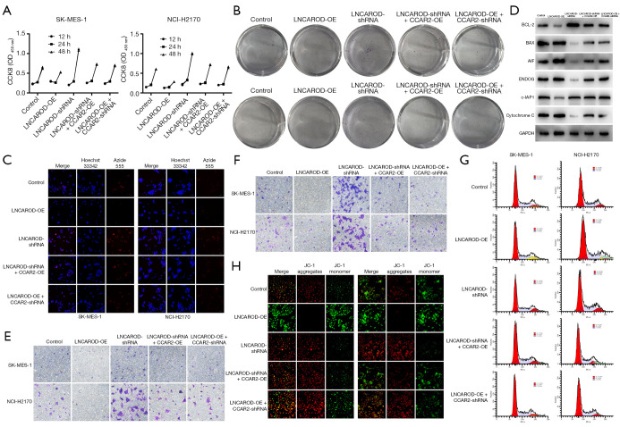
Methods: Using quantitative polymerase chain reaction (qPCR) detection, we determined the expression of LNCAROD in LUSC tissues and cell lines. Cell Counting Kit-8 (CCK-8), EdU (5-ethynyl-2'-deoxyuridine), JC-1 mitochondrial membrane potential, flow cytometry, colony formation, scratch healing, and Transwell assays were conducted, and cell proliferation, migration, and invasion, as well as physiological changes were assessed. The tumorigenicity of LUSC cells was analyzed by in vitro tumor formation in nude mice. Molecular interactions were verified via Western blotting, RNA-protein pull-down assay, RNA binding protein immunoprecipitation (RIP), N6-methyladenosine (m6A)-RIP, and coimmunoprecipitation (Co-IP) analyses.

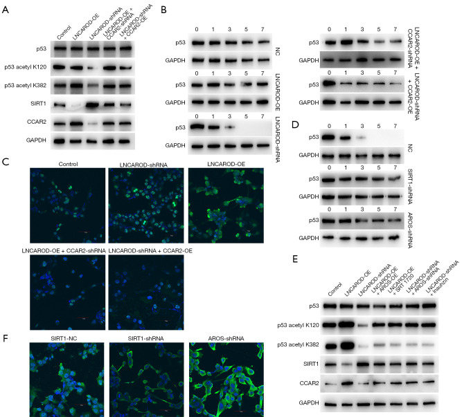
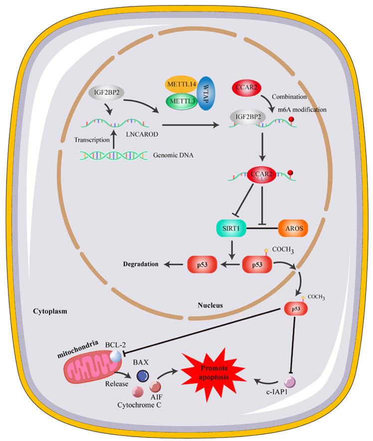
Results: LNCAROD was specifically and highly expressed in LUSC cells and tissues. LNCAROD expression was mediated by IGF2BP2 m6A methylation, which, along with CCAR2, inhibited SIRTI1's acetylation activity. This further induced p53 protein acetylation and promoted the mitochondrial apoptosis of LUSC cells, thereby inhibiting cell proliferation, migration, and invasion.

Conclusions: LNCAROD is specifically highly expressed in LUSC cells and tissues and may be a tumor-suppressor gene. The findings contribute to a deeper understanding of the function of LNCAROD in LUSC, and it may serve as a potential prognostic marker for personalized medical diagnosis in clinical practice.

Keywords: CCAR2; LNCAROD; SIRTI1; lung squamous cell carcinoma (LUSC); m6A methylation.


Conflict of interest statement

Conflicts of Interest: All authors have completed the ICMJE uniform disclosure form (available at https://tlcr.amegroups.com/article/view/10.21037/tlcr-2025-267/coif). R.B. reports that his institution receives funding from Astra Zeneca, Pfizer, and Roche, and that he serves on the Advisory Board of Bayer, Boeringher Ingelheim, EISAI, Lilly, Menarini, and GSK. The other authors have no conflicts of interest to declare.

Figures

Figure 1
Figure 1
Expression of LNCAROD was upregulated in LUSC. (A) The volcano plot shows differentially expressed lncRNAs in LUSC. (B) The heatmap displays the lncRNA profile of lung cancer samples from TCGA. (C) TCGA database analysis of the expression status of LNCAROD in LUSC samples. (D) LUSC expression corresponded to LUSC expression in LUSC and normal tissues. (E) RT-qPCR was performed to detect the expression of LNCAROD in fresh LUSC and adjacent normal tissues. (F) Survival curves were plotted to analyze the relationship between the expression of LNCAROD and overall survival in patients with LUSC. (G) Enrichment analysis of the GSEA gene set. (H) RT-qPCR for the detection of transcripts of LNCAROD in LUSC cell lines and normal cells. (I) RT-qPCR was used to determine the expression of LNCAROD in LUSC cell lines. (J) FISH probes to detect the localization of LNCAROD (200×). ***, P<0.001; ****, P<0.0001; FC, fold change; GSEA, Gene Set Enrichment Analysis; LUSC, lung squamous cell carcinoma; TCGA, The Cancer Genome Atlas; RT-qPCR, real-time quantitative polymerase chain reaction; GSEA, gene set enrichment analysis; FISH, fluorescence in situ hybridization.
Figure 2
Figure 2
LNCAROD exerted inhibitory effects on the proliferation, migration, and invasion of LUSC cells. (A) Establishment of stable LNCAROD-overexpression and -knockdown cell lines. (B) CCK-8 assay was performed to analyze the effects of overexpression and knockdown of LNCAROD on cell proliferation. (C) EdU staining to assess the effect of overexpression and knockdown of LNCAROD on cell proliferation (200×). (D) Clonogenic and crystal violet staining assay to investigate the effect of overexpression and knockdown of LNCAROD on colony formation. (E) Transwell migration and crystal violet staining assay to evaluate the migratory ability of LNCAROD-overexpression or -knockdown LUSC cells (200×). (F) Transwell invasion and crystal violet staining assay to examine the invasive capacity of LNCAROD-overexpression or knockdown LUSC cells (200×). (G) Flow cytometry analysis to detect the effects of LNCAROD overexpression and knockdown on the cell cycle of LUSC cells. CCK-8, Cell Counting Kit-8; LUSC, lung squamous cell carcinoma; NC, negative control; OD, optical density; OE, overexpression.
Figure 3
Figure 3
LNCAROD promoted tumor initiation in vivo. (A) Macroscopic view of nude mice with xenografted tumors. (B) Subcutaneous xenograft tumor body in nude mice. (C) Growth curves of xenografted tumors obtained from control, LNCAROD-overexpression, and LNCAROD-silenced SK-MES-1 cells. (D) Analysis of the weight of xenograft tumors. (E) qPCR detection of LNCAROD expression in tumors. NC, negative control; OE, overexpression; qPCR, quantitative polymerase chain reaction.
Figure 4
Figure 4
IGF2BP2 modulated the stability of LNCAROD through m6A modification. (A) RNA pull-down and Western blot analysis demonstrated the interaction between LNCAROD and IGF2BP2 proteins in SK-MES-1 cells. (B) RIP-qPCR analysis revealed the enrichment of LNCAROD in anti-IGF2BP2 precipitates. (C) MeRIP-qPCR experiments demonstrated an enhanced abundance of LNCAROD upon the silencing of IGF2BP2 in SK-MES-1 and NCI-H2170 cells. (D) SRAMP was used to predict m6A methylation sites on LNCARD. (E) Pulse-chase assays were conducted to assess the stability and expressional dynamics of LNCAROD in SK-MES-1 and NCI-H2170 cells following knockout of IGF2BP2 after treatment with actinomycin D (5 µg/mL) for an indicated duration. IgG, immunoglobulin G; NC, negative control; qPCR, quantitative polymerase chain reaction; RIP, RNA Binding Protein Immunoprecipitation Assay.
Figure 5
Figure 5
LNCAROD enhanced the stability of CCAR2 and facilitated the protein–protein interaction between CCAR2 and AROS. (A) RNA pull-down and Western blot analysis demonstrated the interaction between LNCAROD, CCAR2, and AROS proteins. (B) RIP-qPCR detection indicated the binding of CCAR2 to LNCAROD. (C) Subcellular fractionation and Western blot analysis showed localization of CCAR2 and AROS proteins in the nucleus of SK-MES-1 and NCI-H2170 cells. (D) Schematic representation depicting the full-length transcript of LNCAROD along with its deletion fragments. (E) The association between CCAR2, AROS proteins, and LNCAROD deletion fragments was confirmed through Western blot analysis. (F) Co-IP experiments indicated the coimmunoprecipitation of exogenous and endogenous AROS with CCAR2 in SK-MES-1 cells. (G) Co-IP experiments showed a weakened association between CCAR2 and AROS proteins in SK-MES-1 cells following treatment with RNase A. (H) Co-IP analysis indicated a correlation between CCAR2 and AROS proteins in SK-MES-1 cells transfected with LNCAROD-shRNA or LNCAROD-OE constructs. (I) Effects of silencing or overexpressing CCAR2 on the expression of LNCAROD. (J) Effects of silencing or overexpressing AROS on the expression of LNCAROD. (K) Effects of silencing or overexpressing LNCAROD on the protein expression of CCAR2. (L) Effect of silencing and overexpressing LNCAROD on AROS mRNA expression. (M) Effect of silencing and overexpressing LNCAROD on CCAR2 mRNA expression. (N) Cycloheximide chase analysis of CCAR2 protein levels in CHX-treated SK-MES-1 cells that silenced or overexpressed LNCAROD. (O) MG132 treatment prevented the decrease in CCAR2 protein levels in SK-MES-1 and NCI-H2170 after LNCAROD depletion. (P) Effect of AROS knockdown on CCAR2 protein expression. (Q) Effect of AROS knockdown on CCAR2 mRNA expression. **, P<0.01; ns, no significance. Co-IP, coimmunoprecipitation; NC, negative control; OE, overexpression; qPCR, quantitative polymerase chain reaction; RIP, RNA Binding Protein Immunoprecipitation Assay.
Figure 6
Figure 6
CCAR2 inhibited the activating effect of AROS on SIRT1 and suppressed the deacetylation of p53. (A) LNCAROD inhibited the deacetylation of p53 by CCAR2. (B) Cycloheximide chase analysis clarified the effect of LNCAROD and CCAR2 on the stability of p53 in SK-MES-1 cells. (C) Immunofluorescence staining confirmed the effect of LNCAROD and CCAR2 on the nuclear export of p53 in SK-MES-1 cells (200×). (D) Activation of SIRT1 or overexpression of AROS both facilitated p53 deacetylation. (E) Cycloheximide chase analysis demonstrated the effects of SIRT1 and AROS on p53 stability in SK-MES-1 cells. (F) Immunofluorescence staining confirmed the effect of SIRT1 and AROS on the nuclear export of p53 in SK-MES-1 cells (200×). NC, negative control; OE, overexpression.
Figure 7
Figure 7
Overexpression of CCAR2 rescued the cellular phenotype of LNCAROD knockdown in LNCAROD-shRNA cells, while inhibition of CCAR2 expression rescued the cellular phenotype of overexpression of LNCAROD in LNCAROD-OE cells. (A) The effect of CCAR2 on the viability of SK-MES-1 and NCI-H2170 cells was assessed using CCK-8 assay. (B) The effect of CCAR2 on the proliferation of SK-MES-1 and NCI-H2170 cells was examined through colony formation and crystal violet staining assay. (C) The effect of CCAR2 on the proliferation of SK-MES-1 cells was evaluated using EdU staining (200×). (D) Western blotting for the detection of BCL-2, c-IAP1, BAX, AIF, ENDOG, and cytochrome C protein levels within the cells. (E) Transwell and crystal violet staining assay were conducted to assess the effect of CCAR2 on the migration of LUSC cells (200×). (F) Transwell and crystal violet staining assay were employed to determine the effect of CCAR2 on the invasion of LUSC cells (200×). (G) Flow cytometry analysis was performed to investigate the effect of CCAR2 on the cell cycle in LUSC cells. (H) Mitochondrial membrane potential in LUSC cells, and its alteration due to CCAR2, were detected via JC-1 staining (200×). CCK-8, Cell Counting Kit-8; LUSC, lung squamous cell carcinoma; OE, overexpression.
Figure 8
Figure 8
Schematic of the mechanism. LNCAROD hinders the activation of SIRT1 by facilitating the interaction between CCAR2 and AROS in LUSC cells, thereby suppressing p53 deacetylation. This leads to the acetylation and mitochondrial translocation of p53, thereby activating mitochondrial apoptosis and inducing tumor cell death. Thus, LNCAROD plays a role in suppressing cancer hallmarks in LUSC. LUSC, lung squamous cell carcinoma.

Similar articles

References

    1. Riely GJ, Wood DE, Ettinger DS, et al. Non-Small Cell Lung Cancer, Version 4.2024, NCCN Clinical Practice Guidelines in Oncology. J Natl Compr Canc Netw 2024;22:249-74. 10.6004/jnccn.2204.0023 - DOI - PubMed
    1. Bray F, Laversanne M, Sung H, et al. Global cancer statistics 2022: GLOBOCAN estimates of incidence and mortality worldwide for 36 cancers in 185 countries. CA Cancer J Clin 2024;74:229-63. 10.3322/caac.21834 - DOI - PubMed
    1. Zhong H, Wang L, Zhu X, et al. STAT3 inhibitor Stattic Exhibits the Synergistic Effect with FGFRs Inhibitor Erdafitinib in FGFR1-positive Lung Squamous Cell Carcinoma. J Cancer 2024;15:5415-24. 10.7150/jca.97477 - DOI - PMC - PubMed
    1. Zhang S, Liu L, Shi S, et al. Bidirectional Association Between Cardiovascular Disease and Lung Cancer in a Prospective Cohort Study. J Thorac Oncol 2024;19:80-93. 10.1016/j.jtho.2023.09.004 - DOI - PubMed
    1. Herbst RS, Morgensztern D, Boshoff C. The biology and management of non-small cell lung cancer. Nature 2018;553:446-54. 10.1038/nature25183 - DOI - PubMed

LinkOut - more resources