Review
. 2025 May 2:16:1580108.
doi: 10.3389/fimmu.2025.1580108. eCollection 2025.

Tumor microenvironment and immune-related myositis: addressing muscle wasting in cancer immunotherapy

Affiliations
Review

Tumor microenvironment and immune-related myositis: addressing muscle wasting in cancer immunotherapy

Shuang Ma et al. Front Immunol. .

Abstract

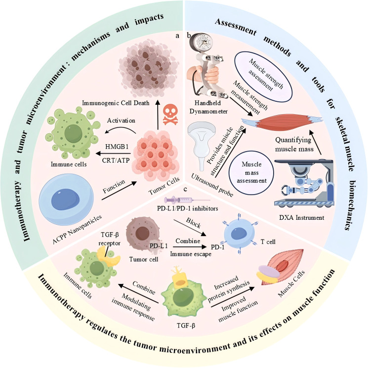
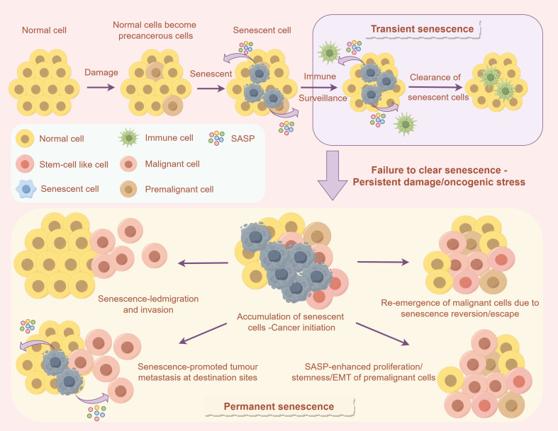
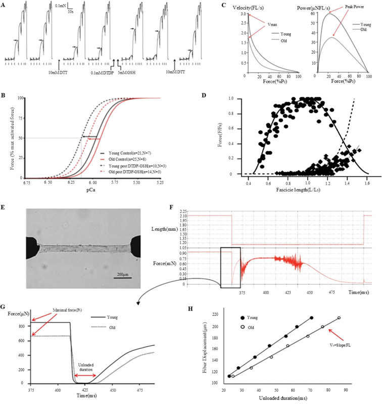
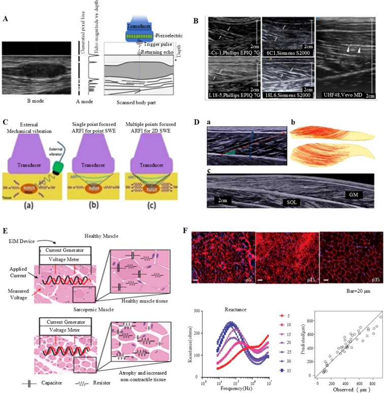
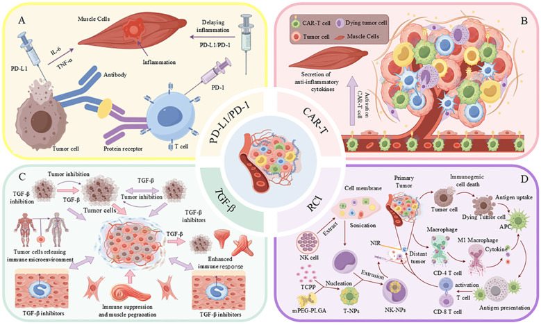
Cancer immunotherapy, which leverages the immune system to target neoplastic cells, has undergone significant transformation in recent. However, immunotherapy may have negative effects on skeletal muscle function, causing muscle wasting and functional decline in cancer patients. In this study, we review the mechanisms by which immunotherapy influences skeletal muscle, focusing on immune-related myositis, inflammation, and metabolic alterations within the tumor microenvironment (TME). The key methodologies, including biomechanical assessment techniques such as electrical impedance myography and ultrasound imaging, are discussed to provide valuable insights into process that maintain muscle integrity and function in patients receiving immunotherapy. Moreover, the dual effects of immunotherapy on tumor suppression and muscle damage are described, revealing the significance of inflammatory cytokines, immune checkpoints, and metabolic disturbances within the TME. Importantly, we propose combination therapies integrating immunotherapy and nutritional interventions or anti-inflammatory interventions as potential approaches for mitigating muscle wasting. This study highlights the need for deeper investigations to optimize immunotherapy and improve its efficacy in preserving muscle health, thereby improving patient outcomes and quality of life.

Keywords: cancer immunotherapy; inflammatory cytokines; muscle wasting; skeletal muscle; tumor microenvironment.


Conflict of interest statement

The authors declare that the research was conducted in the absence of any commercial or financial relationships that could be construed as a potential conflict of interest.

Figures

Figure 1
Figure 1
Immunotherapy modulates the tumor microenvironment and influences skeletal muscle function. (a) Immune activation and tumor cell death mediated by immunogenic signals. (b) Common tools for skeletal muscle strength and mass assessment. (c) Immunotherapeutic blockade of PD-1/PD-L1 and TGF-β pathways enhances immune response and improves muscle function
Figure 2
Figure 2
The influence of cellular senescence on the TME and immune response. The upper section describes how normal cells can enter a temporary senescent state following damage, subsequently being removed through immune surveillance. Conversely, the lower section explains that if senescent cells are not efficiently cleared, they may accumulate as a result of sustained damage or oncogenic stress. This accumulation can result in permanent senescence, thereby facilitating tumor initiation, invasion, and metastasis.
Figure 3
Figure 3
Muscle fiber mechanics and function: effects of DTT, DTDP, and GSH and age-related changes. (A) Investigation of the effects of DTT, DTDP, and GSH on the calcium sensitivity of the contractile apparatus in type II lateral femoral muscle fibers in elderly individuals (80). (B) Analysis of pCa50 and Hill coefficient values derived from Hill curves, based on the mean of individual fit values to the force-pCa relationship, plotted before (control) and after S-glutathionylation treatment for each type II muscle fiber in both young and elderly subjects (80). (C) Examination of the relationship between muscle velocity and force during isotonic contraction (81). (D) Assessment of the in vivo force-length relationship of the human soleus muscle (82). (E) Description of the experimental setup, which includes permeabilized fibers connected to a force transducer and servomotor (81). (F) Outline of the experimental procedure involving the transfer of fibers to an activation solution for relaxation testing (81). (G) Comparative analysis of the maximum force (Po) and no-load duration in type-I fibers of young and elderly subjects (81). (H) Quantification of the no-load duration across various relaxation lengths (81).
Figure 4
Figure 4
Non-invasive assessment of muscle function: ultrasound imaging and electrical impedance myography. (A) Generation of A- and B-mode ultrasonography (98). (B) Longitudinal B-mode ultrasound imaging of the medial gastrocnemius muscle in a healthy volunteer (98). (C) B-mode ultrasound imaging of the gastrocnemius muscle conducted with a linear transducer (98). (D) Comprehensive analysis of the medial gastrocnemius muscle (GM) utilizing ultrasonography: (a) B-mode ultrasound imaging performed with a linear transducer, (b) Diffusion tensor imaging (DTI) fiber reconstruction of the GM, and (c) Extended field-of-view ultrasound imaging of the GM (99). (E) Conceptual illustration of impedance measurements in healthy versus sarcopenic muscles, highlighting increased non-contractile tissue and reduced myocyte size (100). (F) Illustration of employing electrical impedance myography (EIM) data to predict muscle fiber size (100).
Figure 5
Figure 5
Strategies for combined treatment of tumors with immunotherapy and the TME. (A) Tumor cells impede the immune response through the interaction of PD-L1 with PD-1 on T-cells, while simultaneously inducing inflammation via the secretion of TNF-α and IL-6. (B) CAR-T cells contribute to the tumor microenvironment (TME) by secreting anti-inflammatory cytokines and activating M1-type macrophages. (C) Inhibition of TGF-β enhances the immune response and suppresses tumor growth. (D) The activation of immune cells through the application of nanoparticles, such as T-NPs and NK-NPs, in conjunction with near-infrared (NIR) light irradiation, facilitates anti-tumor immune responses.

References

    1. Liu B, Zhou H, Tan L. Exploring treatment options in cancer: Tumor treatment strategies. Signal Transduct Target Ther. (2024) 9:175. doi: 10.1038/s41392-024-01856-7 - DOI - PMC - PubMed
    1. Hsu CY, Pallathadka H, Jasim SA, Rizaev J, Olegovich BD, Hjazi A, et al. Innovations in cancer immunotherapy: A comprehensive overview of recent breakthroughs and future directions. Crit Rev Oncol Hematol. (2025) 206:104588. doi: 10.3390/jcm13216537 - DOI - PubMed
    1. Casagrande S, Sopetto GB, Bertalot G, Bortolotti R, Racanelli V, Caffo O, et al. Immune-related adverse events due to cancer immunotherapy: immune mechanisms and clinical manifestations. Cancers (Basel). (2024) 16:1440. doi: 10.3390/cancers16071440 - DOI - PMC - PubMed
    1. Amy HN, Diana MM, An TN, Eduardo B. Immunotherapy-related skeletal muscle weakness in cancer patients: a case series. Case Reports. (2021) 10:2359–65. doi: 10.21037/apm-20-454 - DOI - PubMed
    1. Jiahua L, Zichao C, Yaqun L, Wenjie Z, JiBiao W, Zhen Z. PD-1/PD-L1 checkpoint inhibitors in tumor immunotherapy. Front Pharmacol. (2021) 12:731798. doi: 10.3389/fphar.2021.731798 - DOI - PMC - PubMed

Substances

LinkOut - more resources