Review
. 2025 May 2:16:1591011.
doi: 10.3389/fimmu.2025.1591011. eCollection 2025.

Comprehending toll-like receptors: pivotal element in the pathogenesis of sepsis and its complications

Affiliations
Review

Comprehending toll-like receptors: pivotal element in the pathogenesis of sepsis and its complications

Wei Wang et al. Front Immunol. .

Abstract

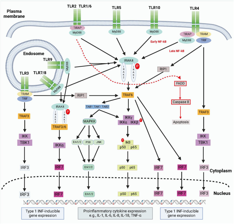
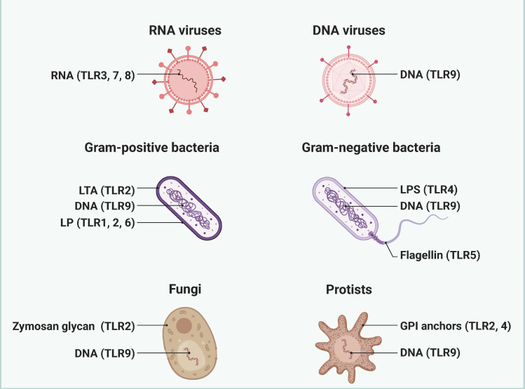
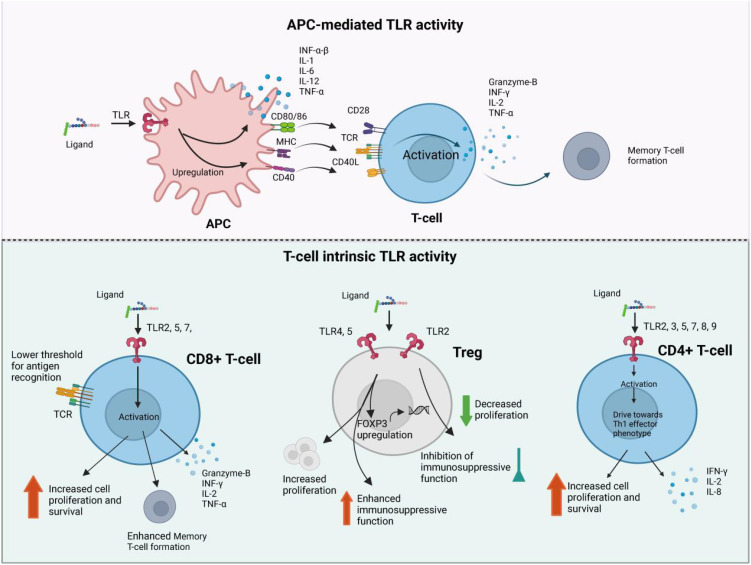
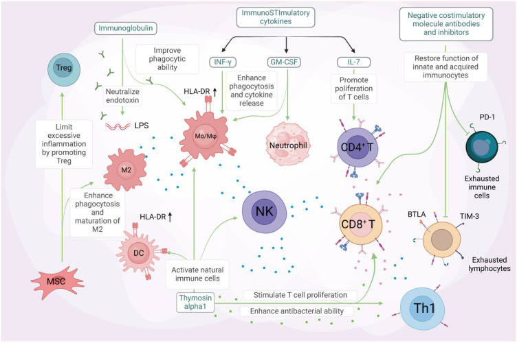
Sepsis, a critical systemic inflammatory response syndrome elicited by pathogenic microorganisms, poses a significant challenge in clinical practice due to its rapid progression and potential for multi-organ failure. This review delineates the intricate roles of Toll-like receptors (TLRs), essential components of the innate immune system, in mediating host responses during sepsis. TLRs recognize pathogen-associated molecular patterns (PAMPs) and damage-associated molecular patterns (DAMPs), thereby initiating signaling cascades that lead to the synthesis of pro-inflammatory cytokines and chemokines. However, the dysregulation of TLR signaling can precipitate a hyper-inflammatory state known as a "cytokine storm," characterized by excessive tissue damage and complications such as Acute Respiratory Distress Syndrome (ARDS) and acute kidney injury (AKI). Several therapeutic strategies targeting TLR pathways are under exploration to mitigate the adverse effects of sepsis. Despite advancements, significant gaps remain, including the need for robust clinical validation and understanding of TLR expression variability among individuals. Future research should focus on elucidating the precise molecular mechanisms governing TLR-mediated responses and developing human-specific therapeutic interventions. This review aims to consolidate current knowledge on TLRs in sepsis, highlighting their dual roles as both defenders against infection and contributors to pathological conditions, thereby informing future therapeutic strategies.

Keywords: damage-associated molecular patterns (DAMPs); inflammation; pathogen-associated molecular patterns (PAMPs); sepsis; sepsis associated complications; toll-like receptors (TLRs).


Conflict of interest statement

The authors declare that the research was conducted in the absence of any commercial or financial relationships that could be construed as a potential conflict of interest.

Figures

Figure 1
Figure 1
The brief summary of TLR signaling pathways. TLRs detect PAMPs or DAMPs on plasma membrane or endosomal membranes. TLRs activate two main pathways: the MyD88-dependent and MyD88-independent pathways. The MyD88-dependent pathway involves MyD88 recruitment and myddosome formation, leading to NF-κB release and proinflammatory cytokine transcription via IKKγ complex activation and MAPK stimulation. The myddosome also promotes IRF5 and IRF7 production, inducing type I IFN gene expression. The MyD88-independent pathway is activated by TLR3 and TLR4, recruiting TRIF to form a complex with TRAF3, TBK1, and IKK. This complex promotes IRF3 nuclear translocation and late-phase NF-κB activation. Both pathways stimulate type I IFN production. Created by biorender.com.
Figure 2
Figure 2
The pathogen ligand targets for different TLRs. Different TLRs are widely recognized as key receptors for recognizing and responding to specific pathogen ligands. Created by biorender.com.
Figure 3
Figure 3
The brief pathogenesis of sepsis. Sepsis pathogenesis entails proinflammatory and anti-inflammatory mechanisms. Proinflammatory and anti-inflammatory responses coexist, but sepsis disrupts their dynamic equilibrium. Initially, there is massive proinflammatory factor secretion, immune cell reprogramming, complement and coagulation system activation, leading to a cytokine storm. Anti-inflammatory cytokines may be released subsequently or concurrently. As sepsis progresses, immune system over-activation results in immune cell depletion and immunosuppression. Sepsis-induced immunosuppression involves immune cell regulation, apoptosis autophagy, endotoxin tolerance, central nervous system changes, and metabolic alterations. Targeting immunosuppression in sepsis can be achieved through these mechanisms. Antigen-presenting cells (APCs), including dendritic cells and macrophages, secrete cytokines such as interferon-α/β (IFN-α/β), interleukin-1 (IL-1), IL-2, and tumor necrosis factor-α (TNF-α). These cells express surface markers CD80/86, MHC class II, and CD40, which facilitate interaction with T-cell receptors (TCR) and co-stimulatory molecule CD28, leading to activation of naïve T-cells. CD8+ cytotoxic T-cells release IFN-γ, IL-2, and TNF-α, alongside the apoptosis-inducing effector molecule Granzyme-B, enhancing their proliferation and cytotoxic activity. CD8+ cytotoxic T-cells release IFN-γ, IL-2, and TNF-α, alongside the apoptosis-inducing effector molecule Granzyme-B, enhancing their proliferation and cytotoxic activity.
Figure 4
Figure 4
Unlocking the potential of immunotherapy in sepsis treatment. Immunomodulatory therapy, combined with anti-inflammatory measures, has been shown to effectively improve the outcome of severe sepsis. Certain immunostimulatory cytokines, including IFN-γ, GM-CSF, and IL-7, have been demonstrated to activate immune cells in sepsis. IFN-γ and GM-CSF enhance phagocytosis and pro-inflammatory cytokine release, as well as the expression of mHLA-DR on antigen-presenting cells (APCs). GM-CSF (middle panel) stimulates neutrophil differentiation and activation through JAK2/STAT5 signaling, while also promoting the survival and proliferation of other myeloid cells (e.g., macrophages and dendritic cells). IL-7 (right panel) drives CD4+ T cell development and homeostasis via JAK1/STAT5 activation, with additional effects on lymphoid lineage cells (including CD8+ T cells and B cell precursors). IL-7, on the other hand, promotes lymphocyte proliferation and inhibits apoptosis. Immunoglobulin, a natural protein, neutralizes endotoxins and enhances the phagocytic ability of monocytes and macrophages. Its administration may benefit septic patients with multidrug-resistant bacterial infections. Thymosin alpha1 activates innate immune cells like dendritic cells (DCs), natural killer (NK) cells, and macrophages. This stimulation leads to T-cell proliferation and enhanced antibacterial effects of Th1 cells. Mesenchymal stem cells (MSCs) facilitate the maturation of M2 macrophages and regulatory T cells, promoting bacterial clearance and reducing excessive inflammation. This alleviates organ damage and ultimately reduces sepsis mortality. Coinhibitory molecule antibodies and antagonists targeting TIM-3, PD-1, BTLA, among others, can restore the function of both innate and adaptive immune cells, reversing the state of immune exhaustion. GM-CSF refers to granulocyte-macrophage colony-stimulating factor, MSC to mesenchymal stem cells, and BTLA to B and T lymphocyte attenuator. Created by biorender.com.

Similar articles

Cited by

References

    1. Zampieri FG, Bagshaw SM, Semler MW. Fluid therapy for critically ill adults with sepsis: A review. JAMA. (2023) 329:1967–80. doi: 10.1001/jama.2023.7560 - DOI - PubMed
    1. Liu D, Huang SY, Sun JH, Zhang HC, Cai QL, Gao C, et al. . Sepsis-induced immunosuppression: mechanisms, diagnosis and current treatment options. Military Med Res. (2022) 9 (1):56. doi: 10.1186/s40779-022-00422-y - DOI - PMC - PubMed
    1. Kellum JA, Formeck CL, Kernan KF, Gomez H, Carcillo JA. Subtypes and mimics of sepsis. Crit Care Clin. (2022) 38:195–211. doi: 10.1016/j.ccc.2021.11.013 - DOI - PubMed
    1. Arora J, Mendelson AA, Fox-Robichaud A. Sepsis: network pathophysiology and implications for early diagnosis. Am J Physiol Regul Integr Comp Physiol. (2023) 324:R613–24. doi: 10.1152/ajpregu.00003.2023 - DOI - PMC - PubMed
    1. Papayannopoulos V. Neutrophil extracellular traps in immunity and disease. Nat Rev Immunol. (2018) 18:134–47. doi: 10.1038/nri.2017.105 - DOI - PubMed

Substances

LinkOut - more resources