. 2025 May 10;53(9):gkaf409.
doi: 10.1093/nar/gkaf409.

Modulation of TTR gene expression in the eye using modified siRNAs

Affiliations

Modulation of TTR gene expression in the eye using modified siRNAs

Jiaxin Hu et al. Nucleic Acids Res. .

Abstract

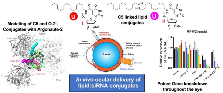
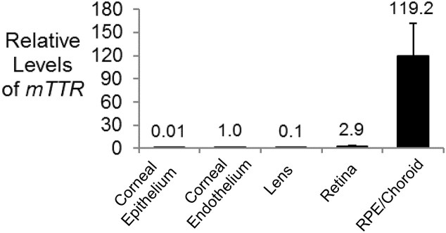
Small interfering RNAs (siRNAs) are a proven therapeutic approach for controlling gene expression in the liver. Expanding the clinical potential of RNA interference requires developing strategies to enhance delivery to extra-hepatic tissues. In this study, we examine inhibiting transthyretin (TTR) gene expression by siRNAs in the eye. Anti-TTR siRNAs have been developed as successful drugs to treat TTR amyloidosis. When administered systemically, anti-TTR siRNAs alleviate symptoms by blocking TTR expression in the liver. However, TTR amyloidosis also affects the eye, suggesting a need for reducing ocular TTR gene expression. Here, we demonstrate that pyrimidine C5- and 2'-O-linked lipid-modified siRNAs formulated in saline can inhibit TTR expression in the eye when administered locally by intravitreal injection. Modeling suggests that length and accessibility of the lipid chains contribute to in vivo silencing. GalNAc-modified siRNAs also inhibit TTR expression, albeit less potently. These data support lipid-modified siRNAs as an approach to treating the ocular consequences of TTR amyloidosis. Inhibition of TTR expression throughout the eye demonstrates that lipid-siRNA conjugates have the potential to be a versatile platform for ocular drug discovery.


Conflict of interest statement

M.M., J.K., and D.D. are employees of Alnylam Pharmaceuticals. V.V.M. is the Paul T. Stoffel/Centex Professor in Clinical Care. D.R.C. is the Rusty Kelley Professor of Biomedical Science. V.V.M., D.R.C., and J.H. hold a patent related to the use of dsRNAs to treat Fuch’s endothelial corneal dystrophy. D.R.C. holds the position of Executive Editor for Nucleic Acids Research and has not peer reviewed or made any editorial decisions for this paper.

Figures

Graphical Abstract
Graphical Abstract
Figure 1.
Figure 1.
(A) Structural representation of C5-lipid, L8 linker, and vinyl phosphonate (VP) modifications used in lipid-modified siRNAs. (B) Duplexes used in these studies. 2′-Fluoro (2′-F) and 2′-O-methyl (2′-OMe) nucleotides are indicated in green and black, respectively. Phosphorothioate (PS) linkages are indicated by orange vertical lines. Uridines linked to lipid at the C5 position are red, and uridines linked to lipid at the 2′-O position are blue.
Figure 2.
Figure 2.
(A) Synthesis of C5 C16-lipid chain containing 2′-O-methyluridine phosphoramidite. (B) Synthesis of C5 C28-lipid chain containing 2′-O-methyluridine phosphoramidite.
Figure 3.
Figure 3.
TTR gene expression after transfection of lipid-modified double-stranded RNAs (dsRNAs) into BNL CL2 murine cells using RNAiMAX as a transfection reagent. (A) TTRa and TTRa–C5–C16. (B) TTRb, TTRb–O2′–C16, and TTRb–C5–C28. (C) TTRa–GalNAc. Error bars are mean with SEM. N = 4 independent replicates.
Figure 4.
Figure 4.
Strategy for ocular injections and analysis. (A) Schematic for the murine eye. (B) Scheme for dissection and tissue analysis. Tissue was harvested 7 days after IVT injection.
Figure 5.
Figure 5.
Relative expression of TTR mRNA in different murine ocular tissues measured by qPCR. Data were normalized relative to levels of murine RPL19. Error bars are mean with SEM. N = 4 independent replicates.
Figure 6.
Figure 6.
Inhibition of TTR gene expression by dsRNAs administered (IVT) at 50 μg per eye. (A) RPE/choroid. (B) Lens capsule. (C) Corneal epithelium/stroma (D) Corneal endothelium. (E) Retina. (F, G) Dose response for TTRa and TTRa–C5–C16. Tissue was harvested 7 days after injection. Data were normalized relative to levels of murine RPL19. Error bars are mean with SEM. N = 4 independent replicates.
Figure 7.
Figure 7.
Inhibition of TTR gene expression in the RPE by dsRNAs TTRb–O2′–C16 and TTRb–C5–C28 at varying concentrations. Tissue was harvested 7 days after IVT injection. Data were normalized relative to levels of murine RPL19. Error bars are mean with SEM. N = 4 independent replicates.
Figure 8.
Figure 8.
Inhibition of TTR gene expression in the RPE by dsRNA TTRa–GalNAc in mouse RPE and liver. (A) Expression levels of TTR in mouse liver. (B) Expression levels in RPE/choroid. Tissue was harvested 7 days after injection of 3 μg (IVT) or 50 μg (SubQ). Data were normalized relative to levels of murine RPL19. Error bars are mean with SEM. N = 4 independent replicates.
Figure 9.
Figure 9.
Computational models of the (A) TTRb–O2′–C16 and (B) TTRb–C5–C28 siRNA duplexes bound to HSA based on the crystal structure of the albumin–myristic acid complex (PDB ID 8RCP). TTRa–C5–C16 can be imagined as a subset of the TTRb–C5–C28 model shown in panel (B). The views are across the major and minor grooves of the RNA duplex (left) and then rotated around the vertical by ca. 90 degrees and more along the helical axis (right). Albumin is depicted in cartoon mode and colored in tan and carbon atoms of fatty acid molecules are green. Bonds of siRNA antisense and sense strands are colored in red and blue, respectively, the 5′-terminal phosphate group of the former is highlighted in black and ball-and-stick mode (bottom left corner of figure panels on the left).
Figure 10.
Figure 10.
Computational models of human Ago2 in complex with an siRNA antisense (AS, black bonds)-sense (S, green bonds) strand duplex with C16 lipid conjugates attached to the O2′ or C5 positions of sense strand residue U6 based on the crystal structure of Ago2 bound to miR-122 opposite a target RNA (PDB ID 6MDZ). Ago2 is depicted in cartoon mode and colored in gray with individual domains labeled (the MID domain is invisible in the background). Carbon atoms of the C16 chain attached to O2′ are colored in cyan. The C16 chain attached to C5 was modeled in two alternative conformations, one emerging from the center of the major groove (carbon atoms colored in pink) and the other wrapping around the backbone of the antisense siRNA to escape the groove (carbon atoms colored in magenta).

Update of

References

    1. Egli M, Manoharan M. Chemistry, structure and function of approved oligonucleotide therapeutics. Nucleic Acids Res. 2023; 51:2529–73.10.1093/nar/gkad067. - DOI - PMC - PubMed
    1. Hofman CR, Corey DR. Targeting RNA with synthetic oligonucleotides: clinical success invites new challenges. Cell Chemical Biology. 2024; 31:125–38.10.1016/j.chembiol.2023.09.005. - DOI - PMC - PubMed
    1. Jadhav V, Vaishnaw A, Fitzgerald K. et al. . RNA interference in the era of nucleic acid therapeutics. Nat Biotechnol. 2024; 42:394–405.10.1038/s41587-023-02105-y. - DOI - PubMed
    1. Nair JK, Willoughby JLS, Chan A. et al. . Multivalent N-acetylgalactosamine-conjugated siRNA localizes in hepatocytes and elicits robust RNAi-mediated gene silencing. J. Am. Chem. Soc. 2014; 136:16958–61.10.1021/ja505986a. - DOI - PubMed
    1. Khan T, Weber H, DiMuzio J. et al. . Silencing myostatin using cholesterol-conjugated siRNAs induces muscle growth. Molecular Therapy - Nucleic Acids. 2016; 5:e342.10.1038/mtna.2016.55. - DOI - PMC - PubMed

Supplementary concepts