. 2025 Nov;77(11):1548-1559.
doi: 10.1002/art.43243. Epub 2025 Jul 21.

Trio Whole Exome Sequencing in Chinese Childhood-Onset Lupus Reveals Novel Candidate Genes

Affiliations

Trio Whole Exome Sequencing in Chinese Childhood-Onset Lupus Reveals Novel Candidate Genes

Jianyang Ma et al. Arthritis Rheumatol. 2025 Nov.

Abstract

Objective: Systemic lupus erythematosus (SLE) is an autoimmune disease in which rare and common gene variants contribute to pathogenesis. Severe sporadic disease in children is often explained by "de novo" variants that can be uncovered by trio sequencing.

Methods: Whole-exome sequencing was performed in 50 Chinese trios with childhood-onset SLE (cSLE). Rare coding variants in SLE-associated genes and all de novo variants were investigated. Gene pathway and expression analysis and interferon-β (IFNβ) luciferase assays were used to predict contribution to disease.

Results: Each proband carried at least one rare variant in an SLE-associated gene, with a median of six per child. At least two probands had monogenic disease, and one-third of probands carried novel or rare variants in genes well accepted to cause monogenic SLE: ACP5, C3, C4A, C4B, DNASE1, IFIH1, NRAS, RNASEH2B, RNASEH2C, and SAMHD1. Probands carried a median of one de novo, rare, coding variant. Intriguingly, although only two de novo variants occurred in genes previously associated with SLE, 12 of the 50 genes were enriched in the top 20 SLE-related pathways and were highly expressed in age-associated B cells and plasma B cells. These genes represent promising candidate lupus genes. Two de novo variants occurring in genes not previously linked to SLE or autoimmunity, DHX8 and ACTR5, enhanced type I IFN signaling.

Conclusion: This study highlights the abundance of lupus-relevant rare gene variants in cSLE, supports frequent contribution of de novo variants to disease, and identifies genes that may constitute novel therapeutic targets of relevance to Chinese patients.


Conflict of interest statement

Declarations of interests

The authors declare no competing interests.

Figures

Figure 1
Figure 1. Patient characteristics and the analysis pipeline.
(A) Ethnicity of the 50 SLE probands. (B) Age and sex distribution. (C) Schematic of the pipeline for rare variant identification. Rare variants are defined as variants with a minor allele frequency (MAF) of <0.005 (gnomAD EAS v4.0). De novo origin of variants was established by comparison with their parents’ sequence data. Putative lupus-associated variants were selected based on filtering rare variants in 49 SLE monogenic genes and 213 GWAS genes. (D) Proportion of variants called by WES. *, splice site variants affecting +1 and +2 nucleotides at the 5′ donor splice site and −1 and −2 residues at the 3′ acceptor splice site. (E) Proportion of patients with different types of variants.
Figure 2
Figure 2. Landscape of rare variants identified by WES in 50 SLE trios.
Rare/novel variants in monogenic SLE genes (orange), GWAS (blue) SLE genes and genes with de novo variants (black), carried by each proband were shown in the pedigree. Paternally inherited gene variants (present in the proband) were listed in the upper left, maternally inherited gene variants (present in the proband) were listed in the upper right, and genes with de novo variants were list below each proband. For genes with multiple variant sites, the number of the variants is shown in parentheses. Monogenic SLE genes with known causal variants: ★; Variants inherited paternally or maternally: #; Compound heterozygous: *; Homozygous: △. ClinVar categories: VUS, Variant of uncertain significance; B, Benign; LB, Likely benign; P/LP, Pathogenic/Likely pathogenic; CI, Conflicting interpretations of pathogenicity. SLE genes validated by expression quantitative trait loci (eQTL) analysis are underlined.
Figure 3
Figure 3. The most frequently mutated genes in 50 SLE trios.
(A) Number of rare variants in known SLE genes (monogenic and GWAS SLE genes) per individual in SLE cohort compared with unaffected parents and controls from the 1000 Genomes Project (1KGP). P values determined by Chi-Square test (*P < 0.05, **** P<0.0001). (B) Most frequently mutated genes in SLE cohort, shown as the proportion of SLE patients and controls carrying rare variants in each indicated SLE-associated gene. Data are based on 64 SLE genes validated by expression quantitative trait loci (eQTL) analysis. Genes with variants occurring at≥2 times the prevalence in controls were shown.
Figure 4
Figure 4. Variants based signaling pathway gene set for enrichment analysis.
All selected genes with rare variants carried by patients were processed with online tool “Metascape” for enrichment analysis. Four pathway analyses were included: GO, KEGG, Reactome and Wiki. (A) The top 20 pathways were automatically generated with the -log10 p value>4 after the enrichment analysis. (B) List of genes of de novo variants that clustered in each of the top 20 pathways. (C) Venn diagram shows the overlap between the 31 genes carrying de novo variants highly expressed in ABCs /plasma cells and the 21 genes carrying de novo variants belonging to the top 20 enriched pathways. *: Taken further for functional validation genes, #: Genes with variants already reported to be pathogenic.
Figure 5
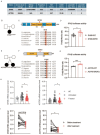
Figure 5. Characteristics of de novo rare variants in two SLE trios.
(A) Bioinformatic predictions of protein damage for each de novo variant based on PolyPhen, SIFT, and CADD score. The gnomAD v4.0 allele frequency, and SNP database (dbSNP) match are shown. EAS = East Asian. (B)(E) Pedigrees of probands carrying de novo rare variants in DHX8 and ACTR5. (C)(F) Schematic representation of protein domain and evolutionary conservation of each residue. (D)(G) IFN-β luciferase assay with indicated wild-type and mutant cDNAs. (H) (I) Left: DHX8 mRNA expression in SLE patients’ PMBCs. HC: healthy control (n=11), untreated patients (n=22), treated patients (n=47), patients before and after treatment (n=15). Right: ACTR5 mRNA expression in SLE patients’ PMBCs. HC: healthy control (n=27), untreated patients (n=22), treated patients (n=47), patients before and after treatment (n=15). P values determined by unpaired t test (D, G), Mann-Whitney t test (H, I). *, P < 0.05; **, P < 0.01; ***, P < 0.001; Graphs depict mean with SD.

References

    1. Cirulli ET, Goldstein DB. Uncovering the roles of rare variants in common disease through whole-genome sequencing. Nat Rev Genet. 2010;11(6):415–25. - PubMed
    1. Lo MS. Monogenic Lupus. Curr Rheumatol Rep. 2016;18(12):71. - PubMed
    1. Brown GJ, et al. TLR7 gain-of-function genetic variation causes human lupus. Nature. 2022;605(7909):349–356. doi: 10.1038/s41586-022-04642-z. - DOI - PMC - PubMed
    1. David C, et al. Gain-of-function human UNC93B1 variants cause systemic lupus erythematosus and chilblain lupus. J Exp Med. 2024;221(8) doi: 10.1084/jem.20232066. - DOI - PMC - PubMed
    1. Xu L, et al. Loss-of-function variants in SAT1 cause X-linked childhood-onset systemic lupus erythematosus. Ann Rheum Dis. 2022;81(12):1712–1721. doi: 10.1136/ard-2022-222795. - DOI - PMC - PubMed

Supplementary concepts