

Additional value of uterine artery Doppler pulsatility index for ultrasound diagnosis of placental site trophoblastic tumor: prospective cohort study
- PMID: 40387112
- PMCID: PMC12209685
- DOI: 10.1002/uog.29235
Additional value of uterine artery Doppler pulsatility index for ultrasound diagnosis of placental site trophoblastic tumor: prospective cohort study
Abstract
Objectives: The ultrasound diagnosis of placental site trophoblastic tumor (PSTT) is challenging owing to a lack of pathognomonic features. Differential diagnosis from other forms of gestational trophoblastic neoplasia (GTN) is critical owing to major differences in prognosis and treatment. Doppler measurement of uterine artery (UtA) pulsatility index (PI) has been proposed for the diagnosis and management of GTN. The aim of this study was to evaluate the added value of UtA-PI Doppler measurement during the standard transvaginal ultrasound (TVS) assessment, in patients with PSTT as compared to those with other GTN.
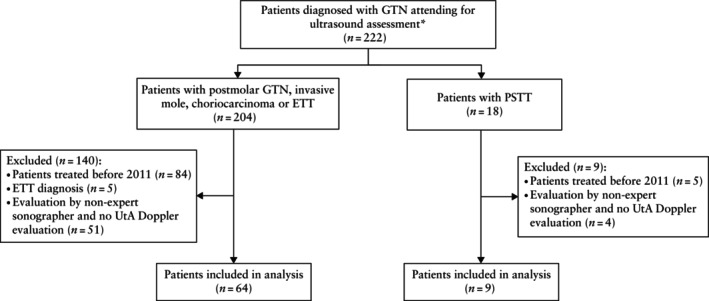
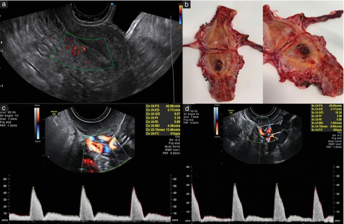
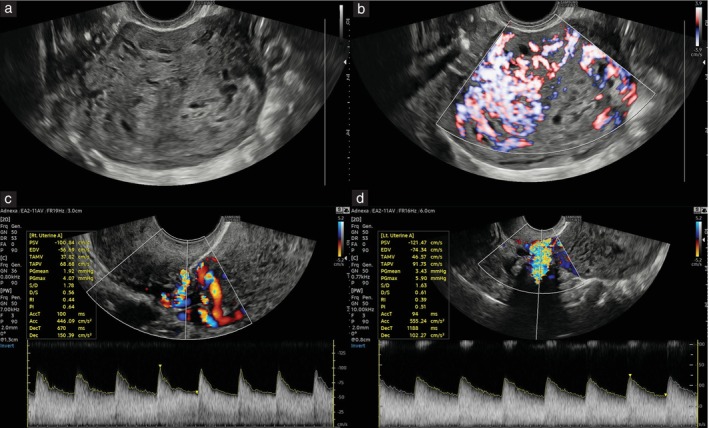
Methods: This was a single-center prospective cohort study involving ultrasound assessment of all GTN cases referred to and treated at the trophoblast unit of San Raffaele Hospital, Milan, Italy, between 2011 and 2023. TVS assessment included: grayscale analysis for the detection of myometrial or endometrial abnormalities, color and power Doppler assessment of lesions with scoring of vascularization, and spectral pulsed-wave Doppler for measurement of mean UtA-PI from the left and right UtAs. Sonographic findings were compared between patients with PSTT and those with other forms of GTN (postmolar, invasive mole or choriocarcinoma), using non-parametric two-tailed statistical analysis.
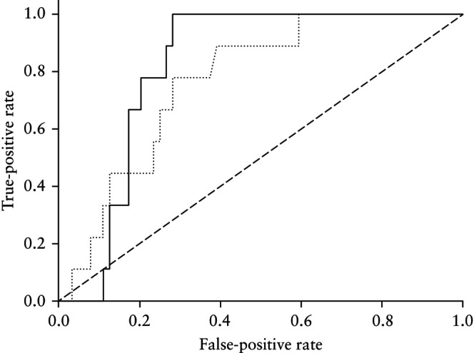
Results: A total of 73 GTN cases were recruited, comprising nine (12.3%) with PSTT and 64 (87.7%) with other GTN. A significant difference was detected between other-GTN and PSTT cases when comparing rates of substantial endometrial vascularity on Doppler (50% vs 0%; P = 0.013) and mean UtA-PI measurements (median, 1.5 (interquartile range (IQR), 1.0-2.4) vs 2.2 (IQR, 1.5-2.7); P = 0.014; area under the receiver-operating-characteristics curve, 0.768 (95% CI, 0.610-0.888)).
Conclusions: This study describes UtA-PI as a novel and effective marker allowing for the ultrasound differentiation of PSTT from other forms of GTN. The significantly higher mean UtA-PI and lower endometrial vascularity observed in PSTT as compared with other GTN suggests a unique vascularization pattern, with a potential role in differential diagnosis and management. © 2025 The Author(s). Ultrasound in Obstetrics & Gynecology published by John Wiley & Sons Ltd on behalf of International Society of Ultrasound in Obstetrics and Gynecology.
Keywords: choriocarcinoma; color‐power Doppler; gestational trophoblastic neoplasia; placental site trophoblastic tumor; uterine artery pulsatility index.
© 2025 The Author(s). Ultrasound in Obstetrics & Gynecology published by John Wiley & Sons Ltd on behalf of International Society of Ultrasound in Obstetrics and Gynecology.
Figures
 ) and combination of presence of endometrial vascularity and mean UtA‐PI (
 ). Area under ROC curve (AUC) for mean UtA‐PI, 0.768 (95% CI, 0.610–0.888). AUC for combined predictors, 0.819 (95% CI, 0.692–0.916).
References
-
- Chawla T, Bouchard‐Fortier G, Turashvili G, et al. Gestational trophoblastic disease: an update. Abdom Radiol (NY). 2023;48(5):1793‐1815. - PubMed
-
- Moutte A, Doret M, Hajri T, et al. Placental site and epithelioid trophoblastic tumours: diagnostic pitfalls. Gynecol Oncol. 2013;128(3):568‐572. - PubMed
-
- Zhao J, Lv WG, Feng FZ, et al. Placental site trophoblastic tumor: a review of 108 cases and their implications for prognosis and treatment. Gynecol Oncol. 2016;142(1):102‐108. - PubMed
Publication types
MeSH terms
LinkOut - more resources
Full Text Sources
Medical
Research Materials
Miscellaneous
