Review

Approach to the Evaluation and Management of Interstitial Lung Abnormalities: An Official American Thoracic Society Clinical Statement

Anna J Podolanczuk et al. Am J Respir Crit Care Med. 2025 Jul.

Erratum in

Abstract

Background: There is growing interest in identifying early stages of interstitial lung disease (ILD) to improve patient outcomes. This document reviews updated evidence on interstitial lung abnormalities (ILAs); provides suggestions for screening, evaluation, and management; proposes criteria for distinguishing ILAs from ILD; and identifies research priorities. Methods: A committee of clinical and methodology experts met by video conference to define ILAs and ILD by consensus and voted on 11 prespecified questions after reviewing synthesized evidence from a systematic literature search. Agreement of ≥70% was required to approve each suggestion. Results: ILA is defined as nondependent bilateral parenchymal abnormalities on computed tomography, including ground-glass opacities or reticulations, lung distortion, traction bronchiectasis, and/or honeycombing involving ≥5% of a lung zone. The updated definition removes the prior exclusion of high-risk populations. ILD is distinguished from ILAs by symptoms (dyspnea/cough) attributable to an interstitial process, abnormal or declining lung function, fibrotic (honeycombing and/or reticulation with traction bronchiectasis involving ≥5% of total lung volume) or progressive imaging abnormalities, and/or specific fibrotic ILD patterns on imaging or pathology. Suggestions include ILA/ILD assessment on imaging acquired for lung cancer screening, screening adults with connective tissue disease and first-degree relatives of patients with familial pulmonary fibrosis, assessing baseline symptoms and pulmonary function among those with ILAs, and monitoring ILAs with chest computed tomography every 2-3 years. Conclusions: This document presents a comprehensive literature review of ILAs with updates to the Fleischner Society ILA definition, establishes a working ILD definition, and provides evidence-based suggestions for ILA evaluation and management.

Keywords: computed tomography; interstitial lung abnormalities (ILA); interstitial lung disease (ILD); pulmonary fibrosis; screening.


Figures
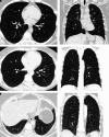
Figure 1.
Figure 1.
Interstitial lung abnormality (ILA) subtypes. (A and B) Nonsubpleural ILA. Axial (A) and coronal (B) images show patchy ground-glass and peribronchovascular opacities predominantly in the upper lung zones with minimal reticular opacities. (C and D) Subpleural nonfibrotic ILA. Axial (C) and coronal (D) computed tomography images show bilateral subpleural reticulations without traction bronchiectasis or honeycombing. (E and F) Fibrotic ILA. Axial (E) and coronal (F) images show bilateral subpleural reticulations with traction bronchiolectasis.
Figure 2.
Figure 2.
Example of interstitial lung disease. Axial (A) and coronal (B) images show bilateral predominantly subpleural reticular opacites with traction bronchiectasis and bronchiolectasis most apparent in the right lung base on the axial image.
Figure 3.
Figure 3.
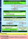
Evaluation and management of interstitial lung abnormality (ILA). Actions shown in darker green boxes indicate recommendations formulated based on prespecified population-intervention-control-outcome (PICO) questions, with the PICO question number shown in parentheses. Actions shown in lighter green boxes indicate additional suggestions that were not the subject of prespecified PICO questions. PICO questions that did not reach consensus are shown in dashed boxes at the bottom of the algorithm in yellow (no recommendation) and red (recommendation against). *Other high-risk populations may be appropriate for screening (e.g., high-risk occupations). Patients at increased risk for lung cancer are recommended to undergo chest computed tomography (CT) screening for lung cancer. We further recommend systematic assessment and documentation of the presence or absence of ILAs/interstitial lung disease (ILD) in smokers who are undergoing lung cancer screening with a chest CT scan. Connective tissue diseases (CTDs) associated with an increased risk of ILD and that qualify for this recommendation include rheumatoid arthritis, systemic sclerosis, polymyositis, dermatomyositis, antisynthetase syndrome, mixed CTD, Sjogren’s disease, undifferentiated CTD, or overlap syndrome. §Including high-resolution CT if not already performed. A multidisciplinary approach is ideal if local resources allow. CTD assessment includes symptom assessment (e.g., skin changes, weakness, arthritis, dry eyes/dry mouth) and physical examination with autoimmune serologies considered when clinically indicated. **Frequency of reassessment depends on risk assessment per Table 4. High-risk features associated with ILA progression include family history of pulmonary fibrosis, older age, smoking history, other inhaled exposures (e.g., occupational vapors, gases, dusts and fumes; air pollution), CTD, presence of the MUC5B promoter variant, leukocyte telomere length below age-adjusted 10th percentile, presence of definite fibrosis on CT (i.e., honeycombing, traction bronchiectasis, or architectural distortion), subpleural fibrotic and subpleural nonfibrotic subtypes, subpleural reticulation, greater extent of imaging abnormalities, and abnormal or borderline FVC, TLC, and DlCO. It may also be appropriate to reassess more frequently for other indications (e.g., lung cancer screening). Implementation depends on local resources. FPF = familial pulmonary fibrosis (defined as at least two genetically related first- or second-degree relatives with fibrotic ILD); IPF = idiopathic pulmonary fibrosis; PFT = pulmonary function test; prevalent ILA = ILA present on baseline and/or follow-up CT scan.

References

    1. Hatabu H, Hunninghake GM, Richeldi L, Brown KK, Wells AU, Remy-Jardin M. et al. Interstitial lung abnormalities detected incidentally on CT: a position paper from the Fleischner Society. Lancet Respir Med . 2020;8:726–737. - PMC - PubMed
    1. Mirza RD, Bolster MB, Johnson SR, Allen A, Jr, Bernstein EJ, Chung JH. et al. Assessing patient values and preferences to inform the 2023 American College of Rheumatology/American College of Chest Physicians interstitial lung disease guidelines. Arthritis Care Res (Hoboken) . 2024;76:1083–1089. - PubMed
    1. Hata A, Schiebler ML, Lynch DA, Hatabu H. Interstitial lung abnormalities: state of the art. Radiology . 2021;301:19–34. - PMC - PubMed
    1. Hata A, Hino T, Yanagawa M, Nishino M, Hida T, Hunninghake GM. et al. Interstitial lung abnormalities at CT: subtypes, clinical significance, and associations with lung cancer. Radiographics . 2022;42:1925–1939. - PMC - PubMed
    1. Salisbury ML, Hewlett JC, Ding G, Markin CR, Douglas K, Mason W. et al. Development and progression of radiologic abnormalities in individuals at risk for familial interstitial lung disease. Am J Respir Crit Care Med . 2020;201:1230–1239. - PMC - PubMed