. 2025 Jun 25;10(6):e0011025.
doi: 10.1128/msphere.00110-25. Epub 2025 May 19.

Mycobacterium tuberculosis bacillus induces pyroptosis in human lung fibroblasts

Affiliations

Mycobacterium tuberculosis bacillus induces pyroptosis in human lung fibroblasts

Takemasa Takii et al. mSphere. .

Abstract

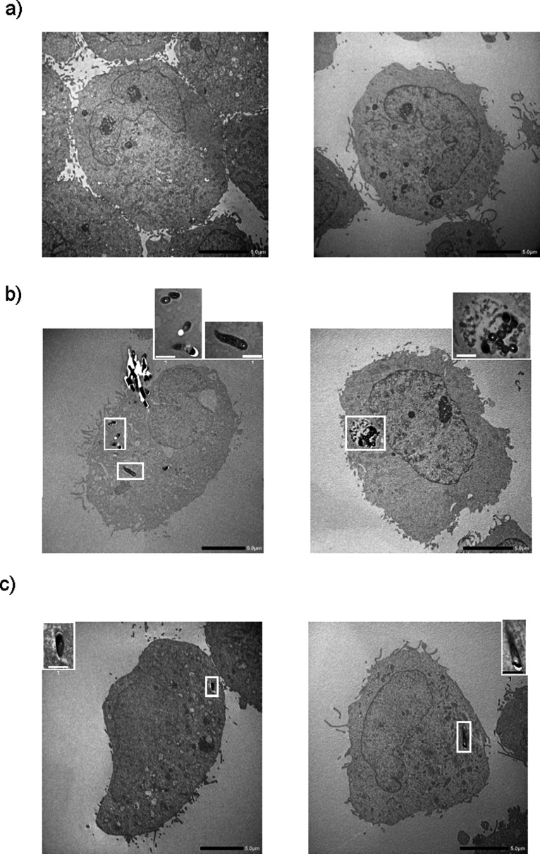
We previously reported that live, but not dead, virulent Mycobacterium tuberculosis (Mtb) H37Rv bacilli induce cell death in human lung fibroblast cell lines, MRC-5, MRC-9, and TIG-1. Here, using two distinct Mtb strains from two different lineages (HN878 lineage 2 and H37Rv lineage 4), we confirmed cell death at day 2 after infection with a device that measures cell growth/cytotoxicity in real time (Maestro-Z [AXION]). Mtb bacilli uptake by the fibroblast was confirmed with a transmission electron microscope on day 2. Expressions of inflammatory cytokines and interleukin (IL)-1β, IL-6, and IL-8 were observed when exposed to live, but not dead bacteria. The cell death of fibroblasts induced by both Mtb strains tested was prevented by caspase-1/4 and NLRP3 inflammasome inhibitors, but not by caspase-3 and caspase-9 inhibitors. Therefore, we classified the fibroblast cell death by Mtb infection as pyroptosis. To investigate the biological and pathological relevance of fibroblast cell death by Mtb infection, we performed dual RNA-Seq analysis on Mtb within fibroblasts and Mtb-infected fibroblasts at day 2. In Mtb bacilli tcrR, secE2, ahpD, and mazF8 genes were highly induced during infection. These genes play roles in survival in a hypoxic environment, production of a calcium-binding protein-inducing cytokine, and regulation of transcription in a toxin-antitoxin system. The gene expressions of IL-1β, IL-6, and IL-8, caspase-4, and NLRP3, but not of caspase-3 and caspase-9, were augmented in Mtb bacilli-infected fibroblasts. Taken together, our study suggests that Mtb bacilli attempt to survive in lung fibroblasts and that pyroptosis of the host fibroblasts activates the immune system against the infection.

Importance: The role of "non-classical immune cells," that is, fibroblasts, epithelial cells, adipocytes, etc., except for the "classical immune cells," that is, macrophages and lymphoid cells, is not well known in the infection of Mtb bacilli. We have previously found that live, but not dead, Mtb bacilli induce cell death in human lung fibroblasts, except in human macrophages and monocytes. The present study reveals that fibroblasts ingest Mtb bacilli the same as macrophages and that in vivo Mtb bacilli within fibroblasts attempt to survive in the host cells, and pyroptosis, including the production of inflammatory cytokines, is induced in the Mtb-infected fibroblasts. Our results suggest that pyroptosis of the host fibroblasts activates the immune system against the infection.

Keywords: Mycobacterium tuberculosis; RNA-Seq; caspase; cytokine; fibroblasts; pyroptosis.


Conflict of interest statement

The authors declare no conflict of interest.

Figures

Fig 1
Fig 1
Time course of cytotoxicity induced by live, but not Mycobacterium tuberculosis (Mtb) bacilli in fibroblasts. Cytotoxicity was measured in real time with Maestro-Z (AXION), which measures the electrical resistance of the fibroblasts adhering to the bottom of an electrode embedded well using CytoView-Z 96-well plate (AXION). Fibroblasts (1 × 10⁴ cells/well) were incubated with live or heat-killed Mycobacterium tuberculosis (Mtb) H37Rv for 96 hours. Initially, 0–100 bacilli were added per fibroblast, (a) MRC-5, (b) MRC-9, and (c) TIG-1, respectively. (d) MRC-5 fibroblasts were incubated with live or heat-killed Mtb HN878 for 96 hours in the same manner. Solid and open shapes indicate the results of live and dead bacilli, respectively. Circle, triangle, and square indicate the number of bacteria added to the fibroblast: 100, 10, 1, respectively. Each experiment was conducted in triplicate. The statistical significance of the difference between the results of each experiment was analyzed by one-way analysis of variance, *P < 0.05, **P < 0.01, ***P < 0.001.
Fig 2
Fig 2
Uptake of Mycobacterium tuberculosis H37Rv bacilli by MRC-5 fibroblasts. MRC-5 fibroblasts (2 × 10⁶ cells/dish) were cultured in a 100 mm diameter tissue culture dish with live Mycobacterium tuberculosis (Mtb) H37Rv (2 × 10⁷ colony-forming units per dish) for the indicated period. After the incubation period, the cells were fixed with glutaraldehyde, and the dehydration steps were performed. After samples were composed with Spurr’s resin, ultrathin sections of each sample were cut to 80 nm thickness with ARTOS 3D (Leica Mikrosysteme GmbH, Vienna, Austria) and stained with uranyl acetate and lead citrate. Transmission electron microscopic examinations were performed with JEM-2100 Plus equipped with a complementary metal-oxide semiconductor CMOS camera with 2,048 x 2,048 pixels. Panels a, b, and c indicate the incubation time of Mtb H37Rv bacilli and MRC-5 fibroblasts on days 1, 2, and 3, respectively. The black bar shows the scale (5.0 µm). The bacilli in the fibroblast are surrounded by a white frame. An enlarged image of the bacilli cut horizontally is shown in the top right corner of panel b. The white bar shows the scale (1.0 µm).
Fig 3
Fig 3
Effects of inhibitors for caspase-1/4, caspase-3, caspase-9, and NLRP3 inflammasome on live Mycobacterium tuberculosis (Mtb) bacilli-induced cytotoxicity to fibroblasts. (a) MRC-5 (1 × 10⁴ cells/well) fibroblasts were seeded on a fibronectin pre-coated CytoView-Z 96-well plate 4 hours before the addition of the inhibitors of caspase-1/4 (VX-765), caspase-3 (PAC-1), caspase-9 (z-LEHD-fmk), and NLRP3 inflammasome (MCC950), and 30 minutes later, the cells were infected with (a) live Mycobacterium tuberculosis (Mtb) H37Rv and Mtb HN878 bacilli (1 × 10⁶ CFU/well). Results are 2 days after incubation with the bacilli. The percentage of inhibition is shown in the panel, and row data are shown in Fig. S3. The error bars for negative numbers were excluded in panel b. MRC-9 and TIG-1 were infected with Mtb H37Rv, and the effects of inhibitors were investigated. Each experiment was performed in triplicates. The statistical significance of the difference between each experiment and the result of incubation with Mtb H37Rv or Mtb HN878 alone were analyzed by one-way analysis of variance, *P < 0.05, **P < 0.01, ***P < 0.001.
Fig 4
Fig 4
mRNA expression of IL-1β, IL-6, and IL-8 in MRC-5 fibroblasts induced by live, but not dead Mycobacterium tuberculosis H37Rv bacilli. MRC-5 fibroblasts (2 × 10⁶ cell/dish) were cultured in a 100 mm diameter tissue culture dish with either live or heat-killed Mycobacterium tuberculosis H37Rv bacilli (2 × 10⁷ bacilli/dish) for the indicated period. mRNA from MRC-5 fibroblasts was extracted with TRIzol reagents (Thermo Fisher Scientific). The mRNA expression of (a) IL-1β, (b) IL-6, and (c) IL-8 was measured by qRT-PCR using the specific primer sets. The statistical significance of the difference between each experiment and the result of incubation with dead bacilli at day 1 were analyzed by one-way analysis of variance, *P < 0.05, **P < 0.01, ***P < 0.001.
Fig 5
Fig 5
The production of IL-1β, IL-6, IL-8, and IL-18 from fibroblasts was induced by live, but not dead Mycobacterium tuberculosis (Mtb) bacilli. Fibroblasts (1 × 10⁴ cells/well) indicated in each panel were cultured in a 96-well plate with either live or heat-killed Mycobacterium tuberculosis (Mtb) H37Rv and/or Mtb HN878 bacilli for 3 days. The amounts of human (a, b) IL-1β, (c, d) IL-6, (e, f) IL-8, and (g, h) IL-18 in the supernatants of fibroblasts were quantified by ELISA. The Med column shows the results without the bacteria. L and D indicated live and dead bacilli, and the numbers, 1, 10, and 100, indicate bacterial number per the fibroblast. Each experiment was conducted in triplicate. The statistical significance of the difference between each experiment and Med result was analyzed by one-way analysis of variance, *P < 0.05, **P < 0.01, ***P < 0.001.
Fig 6
Fig 6
Dual RNA-Seq analysis of in vivo Mycobacterium tuberculosis (Mtb) bacilli. MRC-5 fibroblasts (2 × 10⁶ cells/dish) were co-cultured with Mycobacterium tuberculosis (Mtb) H37Rv bacilli (2 × 10⁷ CFU/dish) for 2 days. According to the reference method (8) for dual RNA-Seq analysis of the Mtb bacilli-infected macrophages, the host cells were solubilized with TRIsol solution. The lysate containing the particles of the in vivo Mtb bacilli was centrifuged, and the supernatant was used for RNA extraction and RNA-Seq analysis of the host cell. Fresh TRIsol solution and zirconia beads were added to the pellet, and bacterial bodies were broken up with a bead beater. After extracting the RNA fractions, the quality of nucleic acid was confirmed, and then RNA-Seq analysis was performed by Macrogen (Kyoto, Japan). (a) Gene ontology (GO) analysis of biological processes (BP), (b) GO analysis of molecular functions (MF), and (c) KEGG pathway analysis. Each experiment was performed in triplicates. The statistical significance of the difference between the result of each experiment and the result of in vitro cultured Mtb bacilli was analyzed by one-way analysis of variance, *P < 0.05, **P < 0.01.
Fig 7
Fig 7
Dual RNA-Seq analysis of Mycobacterium tuberculosis (Mtb) bacilli-infected fibroblasts. The preparation of the RNA fraction is described in the legend to Fig. 6. (a) Gene ontology (GO) analysis of biological processes, (b) GO analysis of molecular functions, (c) GO analysis of cellular components. Each experiment was performed in triplicates.

References

    1. Takii T, Abe C, Tamura A, Ramayah S, Belisle JT, Brennan PJ, Onozaki K. 2001. Interleukin-1 or tumor necrosis factor-alpha augmented the cytotoxic effect of mycobacteria on human fibroblasts: application to evaluation of pathogenesis of clinical isolates of Mycobacterium tuberculosis and M. avium complex. J Interferon Cytokine Res 21:187–196. doi: 10.1089/107999001750133258 - DOI - PubMed
    1. Takii T, Yamamoto Y, Chiba T, Abe C, Belisle JT, Brennan PJ, Onozaki K. 2002. Simple fibroblast-based assay for screening of new antimicrobial drugs against Mycobacterium tuberculosis. Antimicrob Agents Chemother 46:2533–2539. doi: 10.1128/AAC.46.8.2533-2539.2002 - DOI - PMC - PubMed
    1. Yan F, Texter J. 2006. Capturing nanoscopic length scales and structures by polymerization in microemulsions. Soft Matter 2:109–118. doi: 10.1039/b513914g - DOI - PubMed
    1. Tamura M, Tokuda M, Nagaoka S, Takada H. 1992. Lipopolysaccharides of Bacteroides intermedius (Prevotella intermedia) and Bacteroides (Porphyromonas) gingivalis induce interleukin-8 gene expression in human gingival fibroblast cultures. Infect Immun 60:4932–4937. doi: 10.1128/iai.60.11.4932-4937.1992 - DOI - PMC - PubMed
    1. Rastogi N, Labrousse V, de Sousa JP. 1992. Mycobacterial growth and ultrastructure in mouse L-929 fibroblasts and bone marrow-derived macrophages: evidence that infected fibroblasts secrete mediators capable of modulating bacterial growth in macrophages. Curr Microbiol 25:203–213. doi: 10.1007/BF01570720 - DOI - PubMed

LinkOut - more resources