. 2025 May;14(10):e70959.
doi: 10.1002/cam4.70959.

FUT8 Is a Critical Driver of Prostate Tumour Growth and Can Be Targeted Using Fucosylation Inhibitors

Affiliations

FUT8 Is a Critical Driver of Prostate Tumour Growth and Can Be Targeted Using Fucosylation Inhibitors

Kayla Bastian et al. Cancer Med. 2025 May.

Abstract

Background: An unmet clinical need requires the discovery of new treatments for men facing advanced prostate cancer. Aberrant glycosylation is a universal feature of cancer cells and plays a key role in tumour growth, immune evasion and metastasis. Alterations in tumour glycosylation are closely associated with prostate cancer progression, making glycans promising therapeutic targets. Fucosyltransferase 8 (FUT8) drives core fucosylation by adding α1,6-fucose to the innermost GlcNAc residue on N-glycans. While FUT8 is recognised as a crucial factor in cancer progression, its role in prostate cancer remains poorly understood.

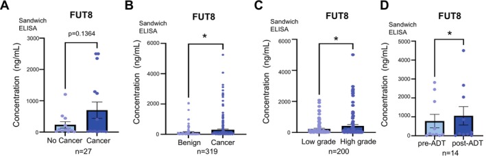
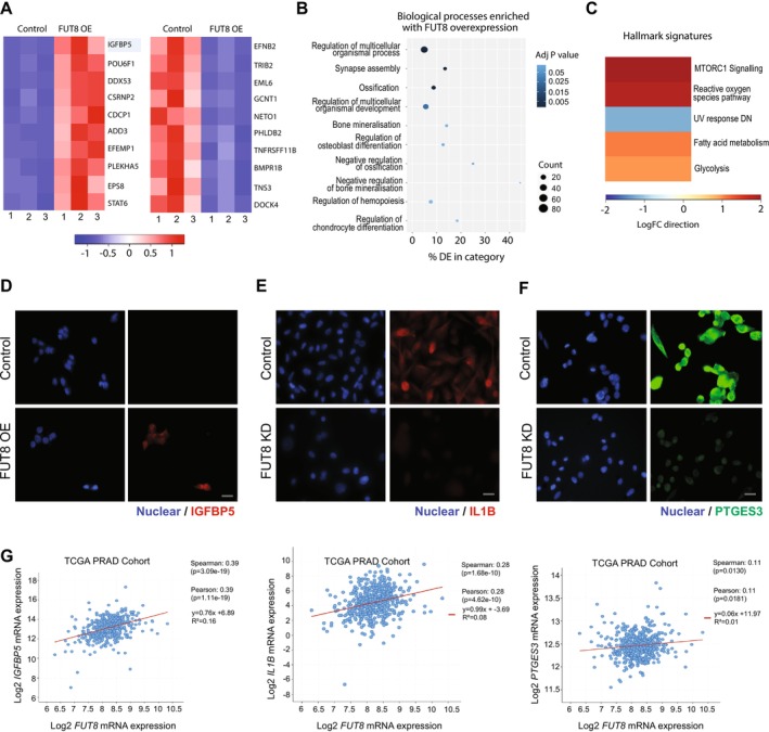
Methods & results: Here, we demonstrate using multiple independent clinical cohorts that FUT8 is upregulated in high grade and metastatic prostate tumours, and in the blood of prostate cancer patients with aggressive disease. Using novel tools, including PhosL lectin immunofluorescence and N-glycan MALDI mass spectrometry imaging (MALDI-MSI), we find FUT8 underpins the biosynthesis of malignant core fucosylated N-glycans in prostate cancer cells and using both in vitro and in vivo models, we find FUT8 promotes prostate tumour growth, cell motility and invasion. Mechanistically we show FUT8 regulates the expression of genes and signalling pathways linked to prostate cancer progression. Furthermore, we find that fucosylation inhibitors can inhibit the activity of FUT8 in prostate cancer to suppress the growth of prostate tumours.

Conclusions: Our study cements FUT8-mediated core fucosylation as an important driver of prostate cancer progression and suggests targeting FUT8 activity for prostate cancer therapy as an exciting area to explore.

Keywords: core fucosylation; fucosylation inhibitors; fucosyltransferase 8 (FUT8); glycans; prostate cancer; therapeutics; tumour growth.


Conflict of interest statement

J.M. and E.S. are shareholders of GlycoScoreDx Ltd. and have filed patents related to this work (GB Patent GB2,594,103 and US Patent App. 17/780,508). J.F.A.P. and E.R. are shareholders of and employed by GlycoTherapeutics B.V. T.J.B. is a shareholder of and scientific advisor of GlycoTherapeutics B.V.; J.F.A.P. and T.J.B. are shareholders of Synvenio B.V. Radboud University and Radboudumc have filed patent applications related to Fucotrim I and Fucotrim II. All other authors declare no conflicts of interest.

Figures

FIGURE 1
FIGURE 1
FUT8 is upregulated in high grade and metastatic prostate tumours. (A–D) FUT8 gene expression levels were detected in clinical samples using real‐time quantitative PCR (RT‐qPCR). (A) FUT8 mRNA levels were significantly higher in prostate cancer relative to benign prostate hyperplasia (BPH) (n = 12, unpaired t‐test, p < 0.01, **). (B) FUT8 mRNA was monitored in a cohort of 33 BPH and 16 prostate cancer samples using real‐time PCR. FUT8 levels were higher in prostate cancer relative to BPH (n = 49, unpaired t‐test, p < 0.01, **). (C) Higher FUT8 expression was also detected in a sub‐group of prostate tumours with ‘metastatic’ biology compared to tumours with a ‘non‐metastatic’ phenotype [67] (n = 20, unpaired t‐test, p < 0.05, *). (D) FUT8 gene expression levels were also significantly increased in metastatic prostate cancer relative to localised disease (n = 20, unpaired t‐test, p < 0.01, **). (E) Immunohistochemistry (IHC) analysis of FUT8 protein levels in a previously published tissue microarray (TMA) [58, 61]. The levels of FUT8 were significantly higher in both Gleason grade 7 tumours (including both 3 + 4 and 4 + 3 tumours) and Gleason grade 8–10 tumours compared to Gleason grade 6 tumours (n = 80, unpaired t test, p = 0.0029 ** and p < 0.0001, ****). Scale bar is 300 μm. (F) Immunohistochemistry analysis of a previously published 125 case TMA [68, 69] to compare FUT8 levels in localised prostate cancer tumours and in prostate cancer tissues presenting with metastasis (all biopsy samples were taken from the primary site). FUT8 levels are significantly higher in metastatic tumours compared to localised tumours (n = 125, unpaired t test, p = 0.0084, **). Scale bar is 200 μm.
FIGURE 2
FIGURE 2
FUT8 protein levels are increased in the blood of patients with aggressive prostate cancer. (A‐D) Detection of FUT8 protein in blood samples from patients with prostate cancer using sandwich ELISA assays. (A) FUT8 levels were 3.02‐fold higher in plasma samples from patients with prostate cancer compared to patients given a no‐cancer diagnosis (n = 27, unpaired t test, p = 0.109). (B) The levels of FUT8 protein were 2.09‐fold higher in plasma samples from men with prostate cancer compared to men diagnosed with BPH (n = 319, unpaired t test, p = 0.0457, *). (C) FUT8 levels were 1.86‐fold increased in serum samples from patients with high grade prostate cancer (Gleason grade 8–9) compared to patients with low grade prostate cancer (Gleason grade 6–7) (n = 200, unpaired t test, p < 0.0218, *). (D) Analysis of FUT8 levels in matched serum samples from 7 men with prostate cancer taken before and after ADT. FUT8 serum levels significantly increase after ADT (n = 14, paired t test, p = 0.047, *).
FIGURE 3
FIGURE 3
Upregulation of FUT8 in prostate cancer cells promotes tumour growth, migration and invasion. (A) Upregulation of FUT8 in CWR22Rv1 cells increases the growth of subcutaneous xenograft tumours. 1 × 107 cells were injected into the flank of CD‐1 nude mice. Tumour size was measured every 3–4 days using callipers. Over 15 days the CWR22V1 tumours with overexpression of FUT8 were 2.23 folf bigger (n = 16, unpaired t test, p = 0.1993). Representative tumour images from each group are shown. (B) Knockdown of FUT8 using shRNA significantly reduces the growth of PC3 tumours in a subcutaneous xenograft model. 3 × 106 PC3 cells were injected into the flank of NMRI mice. Tumour size was measured every 3–4 days using callipers. Over 40 days, the growth of PC3 tumours with knockdown of FUT8 was significantly reduced (n = 12, unpaired t test, p = 0.0055, **). (C‐F) Upregulation of FUT8 in CWR22Rv1 cells promotes cell migration (unpaired t text, p = 0.0092, **) and invasion (unpaired t test, p = 0.0156, *). Knockdown of FUT8 in PC3 cells decreases prostate cancer cell migration (unpaired t test, p = 0.0102, *) and invasion (unpaired t test, p = 0.0113, *). Scale bar is 20 μm.
FIGURE 4
FIGURE 4
FUT8 mediates core fucosylation of N‐glycans in prostate cancer cells. (A,B) Detection of core fucosylated N‐glycans using PhoSL immunofluorescence. (A) PC3 cells with knockdown of FUT8 and have reduced levels of core fucosylated N‐glycans (unpaired t test, p = 0.0227, *) (B) CWR22Rv1 cells with overexpression of FUT8 have increased levels of core fucosylated N‐glycans (unpaired t test, p = 0.0005, ***). Scale bar = 10 μM. Corrected total cell fluorescence (CTCF) indicates a significant decrease in PhoSL binding intensity with FUT8 knockdown, while overexpression of FUT8 significantly increases PhoSL binding intensity. (C) Analysis of FUT8 protein and core‐fucosylated N‐glycans in CWR22Rv1 xenograft tumours (from the experiment shown in Figure 2A) using immunohistochemistry and N‐glycan Matrix‐assisted laser desorption/ionizationmass spectrometry imaging (MALDI‐MSI) to identify core‐fucosylated N‐glycans. Images show the spatial distribution of core fucosylated bi‐antennary N‐glycan (1773.581 m/z), tri‐antennary N‐glycan (1825.5961 m/z) and the complex core fucosylated tetra‐antennary N‐glycan (2190.7632 m/z). EndoF3 cleavage induced a shift of 349.137 amu. Glycan nomenclature: Blue square indicates GlcNAc, yellow circle indicates galactose, green circle indicates mannose, red triangle indicates fucose, and purple diamond indicates sialic acid. Scale bar is 5 mm.
FIGURE 5
FIGURE 5
FUT8 regulates oncogenic genes and proteins in prostate cancer cells. RNA‐sequencing analysis of CWR22Rv1 cells with overexpression of FUT8 identified 381 differentially expressed genes (adjusted p‐value < 0.05, Log2FC 0.58) (Table S2). (A) Heatmap to illustrate the top 10 upregulated and 10 ten downregulated differentially expressed genes. (B, C) Gene Ontology and gene set enrichment analyses of genes regulated by FUT8 revealed CWR22Rv1 cells overexpressing FUT8 have enrichment in ‘ossification’, ‘bone mineralisation’ and ‘regulation of osteoblast differentiation’. (D–F) Validation at the protein level using immunocytochemistry shows (D) IGFBP5 is upregulated when FUT8 is overexpressed in CWR22Rv1 cells and (E, F) Knockdown of FUT8 downregulates IL1B and PTGES3 in PC3 cells. Scale bar is 20 μm. (G) Analysis of the TCGA PRAD cohort shows a significant correlation between the FUT8 gene and levels of IGFBP5, IL1B and PTGES3 in clinical prostate cancer tissue.
FIGURE 6
FIGURE 6
Targeting FUT8‐mediated core fucosylation in prostate cancer with fucosylation inhibitors suppresses tumour growth. (A) CWR22Rv1 cells were subcutaneously injected into the flank of 7‐week‐old CD‐1 nude mice. 7 days prior to implantations mice were randomised to start treatment with either 150 mg/kg fucosylation inhibitor SGN‐2FF or water via oral gavage daily (n = 10 mice/group). Tumour size was measured every 3–4 days using callipers. (B) Tumour volume (mm3) was significantly reduced in the SGN‐2FF treated mice after 21 days (Welch's t‐test for tumour volume on Day 21, p = 0.0034, **). Representative images of tumours are shown. (C) Analysis of CWR22Rv1 xenograft tumours (from experiment shown in Figure 6B) using N‐glycan MALDI‐MSI to identify core‐fucosylated N‐glycans. Images show the spatial distribution of core fucosylated bi‐antennary N‐glycan (1773.581 m/z), tri‐antennary N‐glycan (1825.5961 m/z) and the complex core fucosylated tetra‐antennary N‐glycan (2190.7632 m/z). EndoF3 cleavage induced a shift of 349.137 amu. Glycan nomenclature: blue square indicates GlcNAc, yellow circle indicates galactose, green circle indicates mannose, red triangle indicates fucose, and purple diamond indicates sialic acid. Scale bar is 5 mm. (D) WST‐1 cell proliferation assays show FUT8 overexpression significantly increases the proliferation of CWR22RV1 cells (unpaired t‐test, p < 0.0001, ****), and this is suppressed by treatment with 30 μM of Fucotrim I over 72 h (unpaired t‐test, p < 0.0001, ****). WST‐1 cell proliferation assays also show FUT8 knockdown significantly reduces the proliferation of PC3 cells (unpaired t‐test, p < 0.0001, ****) and by treatment with 30 μM of Fucotrim I for 72 h (unpaired t‐test, p = 0.0012, ***). (E) Colony formation assays show FUT8 overexpression significantly increases the ability of CWR22RV1 cells to survive and grow in colonies over 14 days (unpaired t‐test, p < 0.0001, ****), and this is suppressed by treatment with 30 μM Fucotrim I (unpaired t test, p < 0.0001, ****). PC3 cells with knockdown of FUT8 have reduced colony formation over 14 days (unpaired t‐test, p = 0.019, **). PC3 cells treated with 30 μM Fucotrim I for 14 days have reduced ability to survive and grow in colonies over 14 days (unpaired t‐test, p = 0.0015, ***). (F) Inhibition of fucosylation in TRAMPC2 and RM1 mouse prostate cancer cells Fucotrim I detected using LCA lectin flow cytometry (which recognises core fucosylated N‐glycans [103]. Cells were treated with a range of concentrations of Fucotrim I from 1 nM to 128 μM for 72 h. The mean fluorescence intensities were normalized to a DMSO control. (G) Colony formation assays show treatment with 64 μM Fucotrim significantly reduced cell colony formation for both TRAMPC2 cells (unpaired t‐test, p < 0.0001, ****) and RM1 cells (unpaired t‐test, p < 0.0001, ****) over 7 days.

References

    1. Sung H., Ferlay J., Siegel R. L., et al., “Global Cancer Statistics 2020: GLOBOCAN Estimates of Incidence and Mortality Worldwide for 36 Cancers in 185 Countries,” CA: A Cancer Journal for Clinicians 71, no. 3 (2021): 209–249. - PubMed
    1. Bray F., Laversanne M., Sung H., et al., “Global Cancer Statistics 2022: GLOBOCAN Estimates of Incidence and Mortality Worldwide for 36 Cancers in 185 Countries,” CA: A Cancer Journal for Clinicians 74, no. 3 (2024): 229–263. - PubMed
    1. Archer Goode E., Wang N., and Munkley J., “Prostate Cancer Bone Metastases Biology and Clinical Management (Review),” Oncology Letters 25, no. 4 (2023): 163. - PMC - PubMed
    1. Nuhn P., De Bono J. S., Fizazi K., et al., “Update on Systemic Prostate Cancer Therapies: Management of Metastatic Castration‐Resistant Prostate Cancer in the Era of Precision Oncology,” European Urology 75, no. 1 (2019): 88–99. - PubMed
    1. Mateo J., Fizazi K., Gillessen S., et al., “Managing Nonmetastatic Castration‐Resistant Prostate Cancer,” European Urology 75, no. 2 (2019): 285–293. - PubMed

MeSH terms