

Management of an infectious complication appearing in a transcanine implant: a case report
- PMID: 40388046
- PMCID: PMC12089639
- DOI: 10.1186/s40729-025-00626-6
Management of an infectious complication appearing in a transcanine implant: a case report
Abstract
Background: Maxillary canine impaction is the second most common dental eruption anomaly, affecting approximately 0.2-3% of individuals, with a higher incidence in females. This condition often results in complications such as the misalignment of adjacent teeth, root resorption, and the development of cystic lesions. In some cases, abstention is recommended for impacted canine is kept with the lacteal tooth held on the dental arch. But in the longer term an implant therapy is nevertheless indicated.
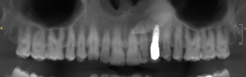
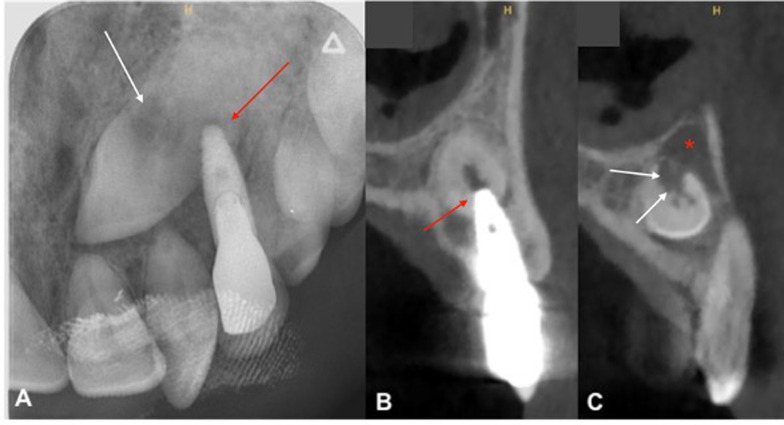
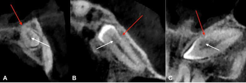
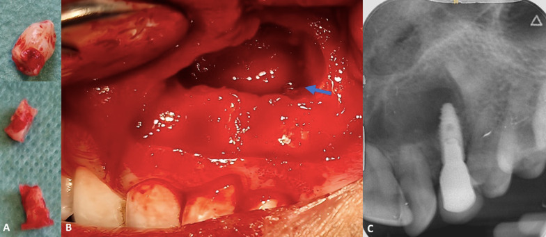
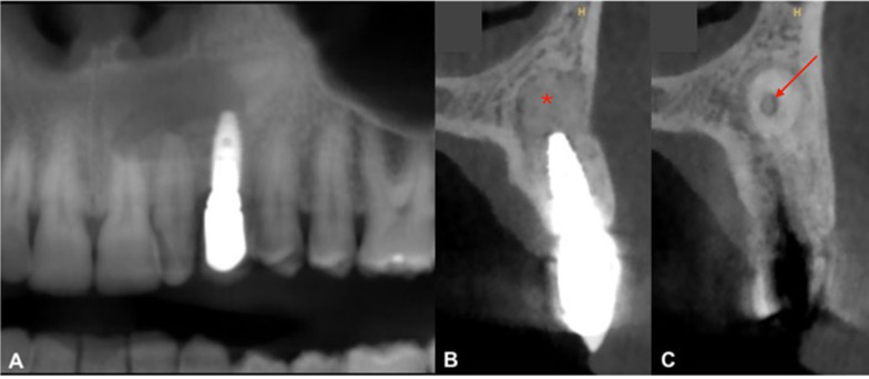
Case presentation: A 42-year-old man presented with persistent swelling and pain in the maxillary region associated with a transcanine implant placed one year ago by his dental practitioner. Imaging assessment showed the implant's apex inserted into the impacted canine which presented a crown and root resorption and was associated to a radiolucency around. In order to preserve implant and reduce morbidity related to a full extraction of the tooth, a coronectomy was performed allowing inflammatory surrounding tissues curettage.
Discussion: This case shows an infectious complication of a transcanine implant and demonstrates an approach for managing these complications while preserving this implant. The coronectomy is a less invasive technique that reduces potential surgical complications and supports healing. A 2-year follow-up revealed complete bone reossification reinforcing the effectiveness of this method in similar clinical scenarios.
Conclusion: This case suggests that coronectomy may be a viable option for managing impacted canines in proximity to implants when complete extraction poses a high risk of complications. However, given the limited number of reported cases and the absence of long-term data, this approach should be considered with caution. Further studies are necessary to better define the indications, long-term outcomes, and potential risks of this technique.
Keywords: Ankylosis; Canine; Coronectomy; Decoronation; Dental implant; Impacted tooth; Transcanine.
© 2025. The Author(s).
Conflict of interest statement
Declarations. Ethics approval and consent to participate: Not applicable. Consent for publication: Informed consent was obtained from the patient in written form. Competing interests: All authors declare that they have no competing interests.
Figures
Similar articles
-
Factors Associated With Successful Surgical Exposure of Impacted Maxillary Canines.J Oral Maxillofac Surg. 2024 Jan;82(1):93-101. doi: 10.1016/j.joms.2023.08.163. Epub 2023 Aug 23. J Oral Maxillofac Surg. 2024. PMID: 37683693
-
Immediate placement of implant into impacted maxillary canine extraction socket.Int J Periodontics Restorative Dent. 2007 Feb;27(1):71-7. Int J Periodontics Restorative Dent. 2007. PMID: 17370664
-
Unconventional implant placement. V: Implant placement through impacted teeth; results from 10 cases with an 8- to 1-year follow-up.Int Orthod. 2015 Jun;13(2):164-180. doi: 10.1016/j.ortho.2015.03.015. Epub 2015 May 23. Int Orthod. 2015. PMID: 26005033 English, French.
-
The Impacted Maxillary Canine in the Adult: A Narrative Review and Implant Treatment Options.J Oral Maxillofac Surg. 2024 Jan;82(1):65-72. doi: 10.1016/j.joms.2023.09.011. Epub 2023 Sep 21. J Oral Maxillofac Surg. 2024. PMID: 37832597 Review.
-
Guidance for the Clinical Management of Impacted Maxillary Canines.Compend Contin Educ Dent. 2021 May;42(5):220-226; quiz 228. Compend Contin Educ Dent. 2021. PMID: 33980019 Review.
References
-
- Becker A, Chaushu S. Etiology of maxillary canine impaction: a review. Am J Orthod Dentofacial Orthop. 2015;148(4):557–67. - PubMed
-
- Bishara SE, Ortho D. Impacted maxillary canines: a review. Am J Orthod Dentofacial Orthop. 1992;101(2):159–71. - PubMed
-
- Izadikhah I, Cao D, Zhao Z, Yan B. Different management approaches in impacted maxillary canines: an overview on current trends and literature. J Contemp Dent Pract. 2020;21(3):326–36. - PubMed
Publication types
MeSH terms
Substances
LinkOut - more resources
Full Text Sources
Miscellaneous
