. 2025 Jun 18;17(24):35066-35079.
doi: 10.1021/acsami.5c03066. Epub 2025 May 19.

Skin Barrier Restoration by Waste-Derived Multifunctional Adhesive Hydrogel Based on Tannin-Modified Chitosan

Affiliations

Skin Barrier Restoration by Waste-Derived Multifunctional Adhesive Hydrogel Based on Tannin-Modified Chitosan

Martina Ferri et al. ACS Appl Mater Interfaces. .

Abstract

The development of multifunctional materials that actively enhance the wound healing process is critically important in addressing clinical and public healthcare challenges. Here, we report a multifunctional hydrogel obtained through physical cross-linking of chitosan and wood tannins for active wound management. Tannins, as polyphenolic wood-waste derivatives, act both as multifunctional additives and cross-linking agents, resulting in a stable and highly swellable hydrogel (>2000%·mg-1). The dressing is produced in the form of a dry and rigid film for easy transportation. After swelling, the material exhibits adequate Young's modulus (∼7 MPa, comparable to the stratum corneum's stiffness), improved flexibility, and suitable adhesion strength to adapt to joint movements. Polyphenolic tannins also provide the material with high antioxidant activity against DPPH radicals (100% RSA), showing potential for preventing complications during the inflammation phase. Moreover, tannins can completely block skin-damaging UV light without significantly altering the material's transparency, thus allowing constant visual wound monitoring. Wound healing investigations on abdominoplasty-derived skin demonstrated that tannins enhance the normal skin barrier restoration process, thereby facilitating the transition toward wound regeneration. This work offers a sustainable strategy for valorizing agri-food waste in a fully biobased material to address active wound management.

Keywords: active wound dressings; chitosan; polyphenols; skin barrier restoration; tannins; waste valorization.


Figures

1
1. Preparation Procedure and Representative Photographs of the Obtained Films at the Investigated Tannin Concentrations (0, 1, 5, and 10 wt %)
1
1
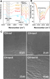
(A) FT-IR (ATR) spectra of CH-ref, CH-tannin films, and tannins (Tan). The main bands are highlighted by gray rectangle and dashed black lines. (B) Highlighted region of FT-IR spectra between 3800 and 2500 cm–1. Black arrow indicates the increase in the −OH broad band intensity with the tannin content. (C) SEM (secondary electrons) of CH-ref, CH-tan1, CH-tan5, and CH-tan10.
2
2
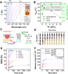
(A) Swelling degree normalized by the mass of the polymer as a function of time. (B) Representative photographs of CH-ref dissolution and its handleability loss compared to CH-tannin’s stability after 24 h of swelling. (C) Gel content (%) and extractable fraction (%) of CH-ref and CH-tannin films with increasing tannin content. (D) Weight loss (%) as a function of time for CH-ref and CH-tannin films at isothermal 37 °C. Inset shows the residue weight (%) and the water evaporation rate (%·min–1) as a function of tannin content in the hydrogel films.
3
3
(A) Representative stress–strain curves obtained from tensile test on dry (warm colors, solid lines) and 5-s swollen samples (cold colors, dashed lines). (B) Young’s modulus (E, MPa) and elongation at break (ε b, %) of the dry and the 5-s swollen films. Arrows highlight the changes in the E and ε b values. (C) Detail on the elongation of CH-tan5 swollen film during the tensile test. (D) Representative adhesion and flexibility test on the skin at the finger joint. Full video is available as Video SV1. (E) Peel adhesion (force/width) over displacement curves of the chitosan and CH-tannin films. A photograph of the test on pig skin is also reported. (F) Adhesion strength (AS) for CH-tannin samples (black dot) as a function of the tannin content and adhesion strength of sticky tape (red dot) as a comparison. Blue, green, and orange shaded areas highlight the ranges of adhesion strength of commercial dressings Tegaderm® (acrylate), BandAid® (acrylate/fabric), and ALGICELL® (alginate), respectively..
4
4
(A) Transmittance of CH-ref and CH-tannin hydrogel films in the UV–vis range. T600, UV–B, and UV–A ranges are highlighted. Inset shows the final transparent appearance of CH-ref and CH-tan10 on a digital wound icon. (B) Transmittance and UV-blocking properties of CH-ref and CH-tannin films. UV–A- and UV–B-blocking activity were calculated by eq . (C) Scheme of the effect of polyphenolic antioxidants on the reactive oxygen species (ROS) produced during the inflammatory stage. (D) Color change of the DPPH solution after the introduction of film samples of CH-ref (0 wt %) and at increasing tannin concentrations (1, 5, 10 wt %). Red arrows indicate the yellow cuvette where 100% of RSA was reached for each sample. (E) Radical scavenging activity (RSA%) as a function of time of the CH-ref and CH-tannin films calculated by using the DPPH method. (F) Tannin release curves in the water medium as a function of time. Inset shows the final tannin release values (24 h) as a function of the tannin content in the films.
5
5
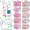
(A) Representative scheme of the organ culture experimental setup. (B) Hematoxylin and eosin (H&E) and (C) claudin 1 expression staining of SDS-perturbed skin following 3 days of contact with CH-ref, CH-tan1, and CH-tan5 patches. Healthy (non-SDS treated) skin and SDS-treated skin without patch application are shown as positive and negative controls, respectively. Black arrows in H&E indicate representative keratinocytes morphology. (D) Claudin 1 expression (at 72 h of incubation) reported as fold increase with respect to SDS sample (8 images for each sample were analyzed). Inset shows time prediction of the fold of increase recovery (at healthy skin levels) of claudin 1 expression for CH-tan1 and CH-tan5. Symbols ** and * represent p-value <0.01 and <0.05, respectively. Data shown are representative of 3 independent experiments.

Similar articles

Cited by

References

    1. Rodrigues M., Kosaric N., Bonham C. A., Gurtner G. C.. Wound Healing: A Cellular Perspective. Physiol. Rev. 2019;99(1):665–706. doi: 10.1152/physrev.00067.2017. - DOI - PMC - PubMed
    1. Carton F., Rizzi M., Canciani E., Sieve G., Di Francesco D., Casarella S., Di Nunno L., Boccafoschi F.. Use of Hydrogels in Regenerative Medicine: Focus on Mechanical Properties. Int. J. Mol. Sci. 2024;25(21):11426. doi: 10.3390/ijms252111426. - DOI - PMC - PubMed
    1. Suarato G., Bertorelli R., Athanassiou A.. Borrowing From Nature: Biopolymers and Biocomposites as Smart Wound Care Materials. Front. Bioeng. Biotechnol. 2018;6:137. doi: 10.3389/fbioe.2018.00137. - DOI - PMC - PubMed
    1. Das P., Manna S., Roy S., Nandi S. K., Basak P.. Polymeric Biomaterials-Based Tissue Engineering for Wound Healing: A Systemic Review. Burns Trauma. 2023;11:tkac058. doi: 10.1093/burnst/tkac058. - DOI - PMC - PubMed
    1. Mao G., Tian S., Shi Y., Yang J., Li H., Tang H., Yang W.. Preparation and Evaluation of a Novel Alginate-Arginine-Zinc Ion Hydrogel Film for Skin Wound Healing. Carbohydr. Polym. 2023;311:120757. doi: 10.1016/j.carbpol.2023.120757. - DOI - PubMed

LinkOut - more resources