. 2025 Jul 9;15(7):jkaf111.
doi: 10.1093/g3journal/jkaf111.

Site-to-site mutational dissection of fission yeast cohesin reveals its dynamics

Affiliations

Site-to-site mutational dissection of fission yeast cohesin reveals its dynamics

Qi Wei et al. G3 (Bethesda). .

Abstract

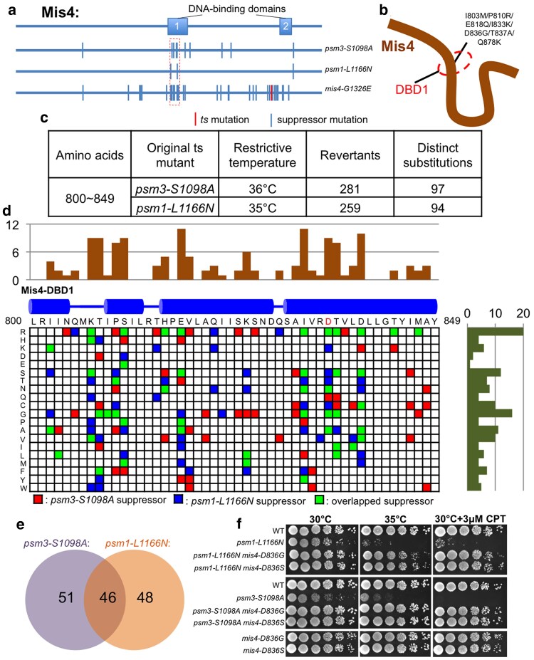
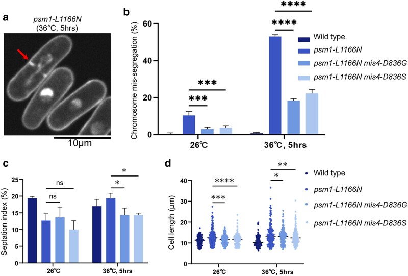
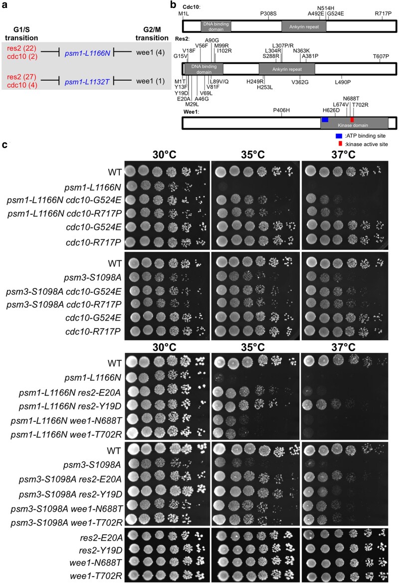
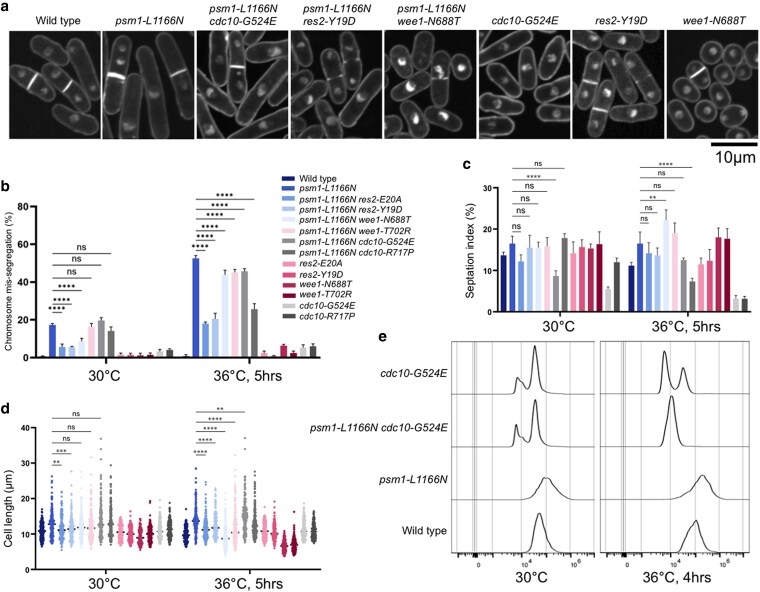
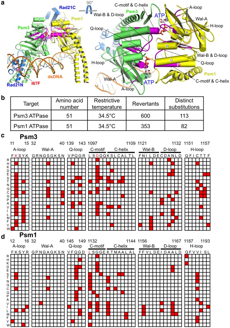
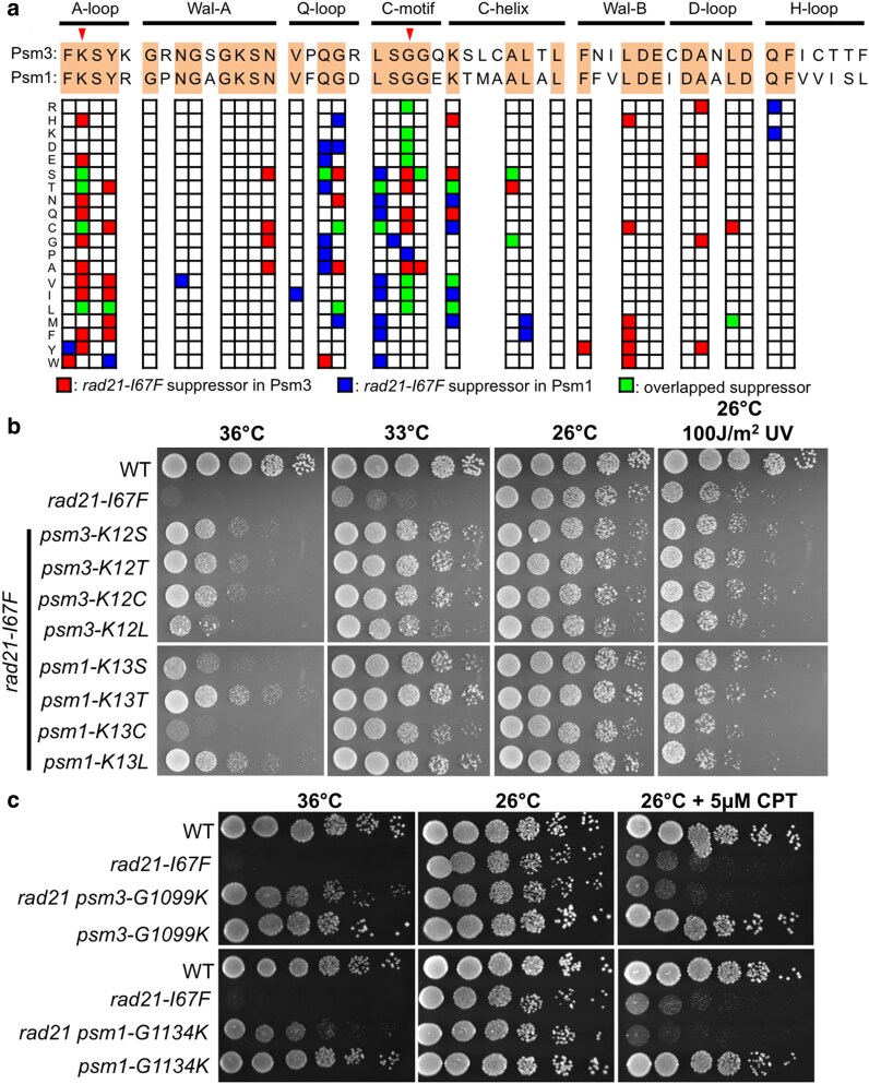
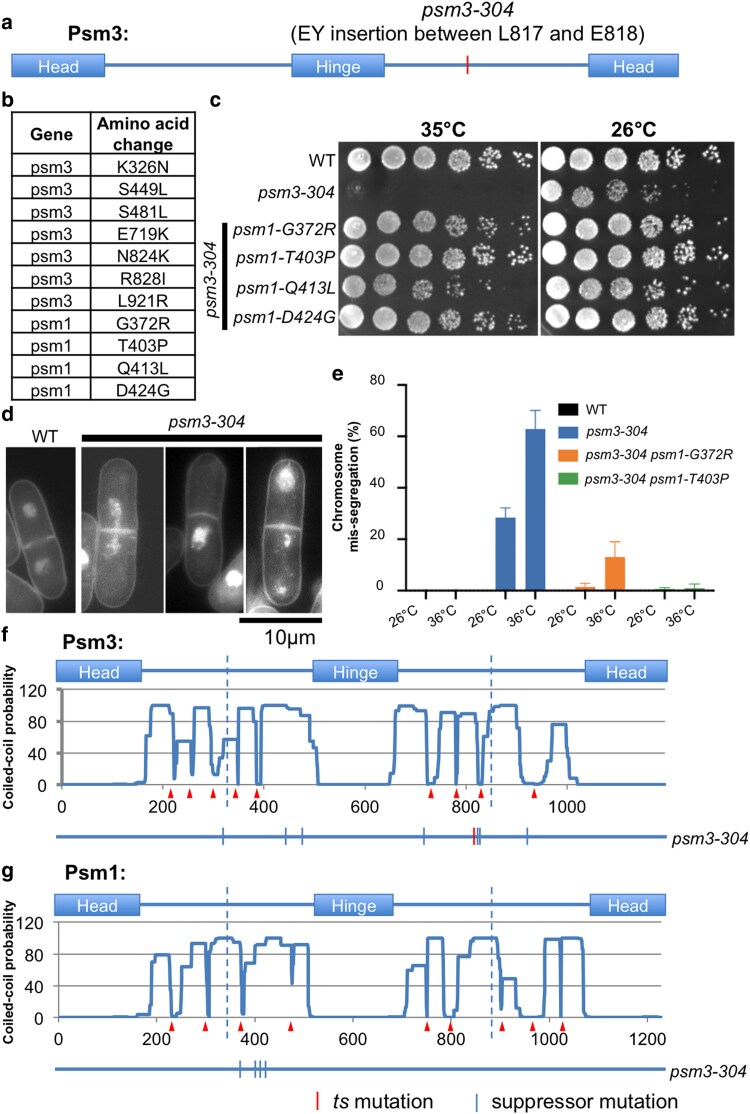
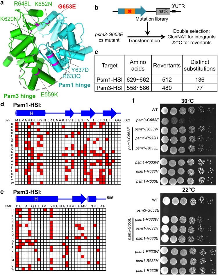
Cohesin is a heteropentameric protein complex that holds sister chromatids together from S phase to anaphase. Its 2 structural maintenance of chromosome subunits form a heterodimer, consisting of an ATPase head domain and a hinge domain connected by long coiled coils. Kleisin subunit associates with the head. Here, using Schizosaccharomyces pombe, we genetically dissected cohesin dynamics based on the relationship between the mutations causing temperature-sensitive and their suppressor mutations. First, we identified suppressor mutations that could rescue the lethality caused by cohesin ATPase mutations. Mutations in the DNA-binding domain of cohesin loader Mis4, or in cell-cycle genes encoding MBF transcription factor complex or Wee1 kinase, rescued both Psm1 and Psm3 ATPase mutants. Then, we performed targeted mutagenesis in both ATPase domains for single-amino-acid substitutions that can rescue the lethality of a kleisin ts mutant at restrictive temperature. Comparison of mutations obtained in Psm1 and Psm3 ATPase domains revealed that analogous mutations in the 2 ATPase domains were frequently observed. Last, suppressors of a coiled-coil mutation were mapped in coiled coils, indicating that proper folding of coiled coils is critical for cohesin functions. Suppressors of a hinge interface mutation are frequently located at the other hinge interface, indicating that the 2 cohesin hinge interfaces work collaboratively in hinge-hinge interactions. Overall, genetic dissection of the relationship between cohesin lethal mutations and their suppressor mutations reflects cohesin dynamics in vivo.

Keywords: cell cycle; chromosome segregation; cohesin; fission yeast; suppressor screen.


Conflict of interest statement

Conflicts of interest: The authors declare no conflicts of interest.

Figures

Fig. 1.
Fig. 1.
Suppressors of cohesin ATPase ts mutants mapped in Mis4. a) Localization of suppressor mutations of psm1-L1166N, psm3-S1098A, and mis4-G1326E identified in Mis4. Amino acids: 800∼849 were highlighted by a dashed rectangle. b) Suppressor mutations located in the DBD 1 (Mis4-DBD1; amino acids: 800∼849) of Mis4. c) Summary of the targeted mutagenesis for identification of single-amino-acid substitutions that can rescue the temperature sensitivity of the cohesin ATPase ts mutants (psm1-L1166N and psm3-S1098A). d) The single-amino-acid substitutions (the colored squares in the matrix) identified as suppressors of the psm1-L1166N and/or psm3-S1098A ts mutants. e) Mutational overlap between the psm1-L1166N suppressors and the psm3-S1098A suppressors in Mis4-DBD1. f) Spot test analysis.
Fig. 2.
Fig. 2.
Phenotypic suppression of a cohesin ATPase mutant by mutations in Mis4. a) DAPI staining followed by microscopic observation for psm1-L1166N mutant cells cultured at restrictive temperature 36°C for 5 h. A cell with chromosome mis-segregation was indicated by an arrow. b) Frequency of chromosome mis-segregation events calculated by counting 200 mitotic cells. c) Septation index calculated by counting number of cells having septation in 200 cells. d) Cell length was measured using the ImageJ software.
Fig. 3.
Fig. 3.
Suppressors of cohesin ATPase mutants in Res2-Cdc10 and Wee1. a) Suppressor mutations of the psm1-L1166N and psm3-S1098A mutants were identified in res2, cdc10, and wee1. b) Mutation localizations in Cdc10, Res2, and Wee1. Suppressor mutations of psm1-L1166N in Res2 were shown above the Res2 protein sequence, while suppressor mutations of psm3-S1098A in Res2 were shown below the Res2 protein sequence. c) Spot test analysis.
Fig. 4.
Fig. 4.
Phenotypic suppression of a cohesin ATPase mutant by mutations in Cdc10, Res2, and Wee1. a) Microscopic observation after DAPI staining of the mutants cultured at 36°C for 5 h. b) Frequency of chromosome mis-segregation events calculated by counting 200 mitotic cells. c) Septation index calculated by counting number of cells having septation in 200 cells. d) Cell length was measured using the ImageJ software. e) Flow cytometry analysis.
Fig. 5.
Fig. 5.
Mutations in cohesin ATPase domains rescued rad21. a) Structural view of cohesin head and locations of the conserved motifs in the 2 cohesin ATPase domains. A cohesin structure (PDB code: 6YUF) was used for presentation. Localization of the Rad21-I67F mutation was indicated too. b) Summary of the genetic screen, targeted to the 2 cohesin ATPase domains, for suppressor mutations that alleviated the premature cohesin releasing phenotype in rad21-I67F. c and d) Data matrices presenting single-amino-acid substitutions identified in Psm3 ATPase domain (c) or Psm1 ATPase domain (d) that rescued the temperature sensitivity of rad21-I67F at restrictive temperature.
Fig. 6.
Fig. 6.
Analogous mutations identified in the 2 cohesin ATPase domains. a) Suppressor mutations of the rad21-I67F ts mutant, identified in the 2 cohesin ATPase domains, were presented in 1 data matrix. Locations of Psm3-K12 (and its corresponding amino acid Psm1-K13) in A-loop and Psm3-G1099 (and its corresponding amino acid Psm1-G1134) in C-motif were indicated by arrowheads. b) Spot test results of the analogous suppressor mutations at the conserved lysine in A-loop (Psm3-K12 corresponds to Psm1-K13). c) Spot test results of the analogous suppressor mutations at the conserved glycine in C-motif (Psm3-G1099 corresponds to Psm1-G1134).
Fig. 7.
Fig. 7.
Suppressors of a Psm3 coiled-coil ts mutant identified in Psm1 and Psm3 coiled coils. a) The psm3-304 ts mutant contains a 2 amino acids (“EY”) insertion in the Psm3 coiled coil between L817 and E818. b) Suppressor screen for the psm3-304 mutant at restrictive temperature (37°C), followed by next-generation sequencing, identified mutations in Psm1 and Psm3. c) Spot test analysis. d) DAPI staining followed by microscopic observation for psm3-304 mutant cells cultured at restrictive temperature 36°C for 5 h. e) Frequency of chromosome mis-segregation events calculated by counting number of cells exhibiting chromosome mis-segregation in 200 mitotic cells. f and g) Localization of the psm3-304 suppressor mutations in the Psm3 and Psm1 protein sequences. The arrowheads demonstrated the positions of discontinuities (or breaks) that were predicted in the Psm1 and Psm3 coiled coils. The vertical dashed lines indicated the middle positions of the coiled coils.
Fig. 8.
Fig. 8.
Suppressors of a Psm3 hinge interface mutant identified in Psm1 and Psm3 hinges. a) Suppressor mutations of a Psm3 hinge interface mutant, psm3-G653E, that were identified by next-generation sequencing. Revertants were isolated at restrictive temperature 22°C. b) Strategy of targeted mutagenesis, transformation, and revertant screening. c) Summary of the suppressor screen. d and e) Data matrices presenting the single-amino-acid substitutions identified in the Psm1 hinge domain d) or Psm3 hinge domain e) that rescued the cold sensitivity of psm3-G653E. f) Spot test analysis.

Similar articles

References

    1. Anderson DE, Losada A, Erickson HP, Hirano T. 2002. Condensin and cohesin display different arm conformations with characteristic hinge angles. J Cell Biol. 156(3):419–424. doi: 10.1083/jcb.200111002. - DOI - PMC - PubMed
    1. Arumugam P, Gruber S, Tanaka K, Haering CH, Mechtler K, Nasmyth K. 2003. ATP hydrolysis is required for cohesin's association with chromosomes. Curr Biol. 13(22):1941–1953. doi: 10.1016/j.cub.2003.10.036. - DOI - PubMed
    1. Bauer BW, Davidson IF, Canena D, Wutz G, Tang W, Litos G, Horn S, Hinterdorfer P, Peters JM. 2021. Cohesin mediates DNA loop extrusion by a “swing and clamp” mechanism. Cell. 184(21):5448–5464.e22. doi: 10.1016/j.cell.2021.09.016. - DOI - PMC - PubMed
    1. Bernard P, Schmidt CK, Vaur S, Dheur S, Drogat J, Genier S, Ekwall K, Uhlmann F, Javerzat JP. 2008. Cell-cycle regulation of cohesin stability along fission yeast chromosomes. EMBO J. 27(1):111–121. doi: 10.1038/sj.emboj.7601955. - DOI - PMC - PubMed
    1. Biegert A, Mayer C, Remmert M, Soding J, Lupas AN. 2006. The MPI bioinformatics toolkit for protein sequence analysis. Nucleic Acids Res. 34(Web Server):W335–W339. doi: 10.1093/nar/gkl217. - DOI - PMC - PubMed

MeSH terms

LinkOut - more resources