. 2025 May 19;20(5):e0322880.
doi: 10.1371/journal.pone.0322880. eCollection 2025.

Evolution and classification of Ser/Thr phosphatase PP2C family in bacteria: Sequence conservation, structures, domain distribution

Affiliations

Evolution and classification of Ser/Thr phosphatase PP2C family in bacteria: Sequence conservation, structures, domain distribution

Hang Li et al. PLoS One. .

Abstract

Serine/threonine kinases (STKs) and serine/threonine phosphatases (STPs) are widely present across various organisms and play crucial roles in regulating cellular processes such as growth, proliferation, signal transduction, and other physiological functions. Recent research has increasingly focused on the regulation of STKs and STPs in bacteria. STKs have been well studied, identified and characterized in a variety of bacterial species. However, the role of STPs in bacteria remains less understood, and the number of proteins characterized is limited. It has been found that most of the STPs characterized in bacteria were Mg2+/Mn2+ dependent 2C protein phosphatases (PP2Cs), but the evolutionary relationship and taxonomic distribution of bacterial PP2C phosphatases were still not fully elucidated. In this study, we utilized bacterial PP2C phosphatase sequences from the InterPro database to perform a phylogenetic analysis, categorizing the family into five groups. Based on this classification, we examined the evolutionary relationships, species distribution, sequence and structural variations, and domain distribution characteristics of bacterial PP2C phosphatases. Our analysis uncovered evidence of a common evolutionary origin for bacterial PP2C phosphatases. These findings advance the understanding of PP2C phosphatases, offering valuable insights for future functional studies of bacterial serine/threonine phosphatases and aiding in the design of targeted therapeutics for pathogenic bacteria.


Conflict of interest statement

The authors have declared that no competing interests exist.

Figures

Fig 1
Fig 1. Phylogenetic tree of bacterial PP2C phosphatases.
The Bootstrap value, obtained from 1000 repetitions, shows nodes with > 90% support as a light blue five-pointed star. The orange, yellow, blue, green, and gray circles on the outer layers of the evolutionary tree represent groups I, II, III, IV, and V, respectively.
Fig 2
Fig 2. The distribution of phylum levels of PP2C phosphatases in five groups.
The distribution of PP2C phosphatases (PF13672) in eukaryotes, archaea, and bacterial phyla was illustrated by a string diagram. Each chord connected different subtypes and phyla, with the color of the chord representing different phyla. The color map on the right represented eukaryotes, archaea, and the total number of the top 17 bacterial phyla. In the upper part of the string diagram, the blue, light green, green, purple, and pink circles represented Groups I, II, III, IV, and V, respectively. The thickness of the chord reflected the relative abundance or number of species.
Fig 3
Fig 3. Sequence logos for conserved motifs in bacterial PP2C phosphatases.
The height of different amino acids represents repeatability. The scale bar at the bottom indicates the length of the motif protein sequence. (A) Sequence conservation and motifs distribution in the core catalytic domain of 3573 candidate sequences. Eleven motifs were labeled on the sequence logo. Conserved Asp in Motifs 1, 2, 8, and 11 were marked with red triangles, while those in Motif 5 were marked with purple triangles. Conserved Gly and Thr residues are indicated by blue and yellow triangles, respectively. (B) Sequence logos for conserved motifs in PP2C phosphatases of five groups, respectively. Conserved Arg residues were labeled with black triangles in motifs. The conserved “DN” sequence and “DD” sequence in Motif 11 were marked with blue and black short lines, respectively.
Fig 4
Fig 4. Comparison of bacterial PP2C phosphatase structures.
(A) Known and predicted structures of representative proteins from five distinct groups: Q8VQA1 (PDB ID: 2PK0, shown in golden) from Streptococcus agalactiae, P9WHW5 (PDB ID: 1TXO, shown in blue) from Mycobacterium tuberculosis, A0A1L7GEB2 (AlphaFold ID: AF-A0A1L7GEB2-F1, shown in purple) from Streptomyces sp. TN58, A0A7V6HC85 (AlphaFold ID: AF-A0A7V6HC85-F1, shown in green) from Clostridiaceae bacterium, and P76395 (AlphaFold ID: AF-P76395-F1, shown in coralline) from Escherichia coli. Metal ions were shown as colored spheres. (B) Structural superposition of five representative proteins. The five representative protein structures were superimposed using ChimeraX software, and the amino acid residues from the active center were labeled based on the sequence alignment results. Asp, Asn, Gly, Ser and Thr residues were labeled with red, orange-red, orange, green and yellow respectively. (C) Flap subdomain and active center of PP2C phosphatases. The flap subdomain is a frontal extension of alpha-helices outside the catalytic domain. In amplified active centers, motifs involved in the formation of active centers were labeled.
Fig 5
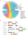
Fig 5. Domain arrangements in bacterial PP2C phosphatases.
(A) The distribution of domains across different bacterial phyla was visualized using string graphs. The color map on the right represented the total number of the top 12 bacterial phyla. The thickness of the chord reflected the relative abundance or number of species in different domain types. (B) Domain arrangements in bacterial PP2C phosphatases. The domain annotation results of the proteins with uniprot IDs: A0A948RMV2, A0A3N6HDR9, A0A2D5QNQ7, N6ZYJ5, A0A0P9RY45, A0A0M1JLJ3, A0A2N2UMZ8, and A0A2S6GKU5. The PP2C, zinc_ribbon, DZR, Pkinase, TPR, cNMP_binding, FHA, MerR, PASTA domains are represented by different colored rectangles.

Similar articles

Cited by

References

    1. Macek B, Forchhammer K, Hardouin J, Weber-Ban E, Grangeasse C, Mijakovic I. Protein post-translational modifications in bacteria. Nat Rev Microbiol. 2019;17(11):651–64. doi: 10.1038/s41579-019-0243-0 - DOI - PubMed
    1. Kyriakis JM. In the beginning, there was protein phosphorylation. J Biol Chem. 2014;289(14):9460–2. doi: 10.1074/jbc.R114.557926 - DOI - PMC - PubMed
    1. DeVinney R, Steele-Mortimer O, Finlay BB. Phosphatases and kinases delivered to the host cell by bacterial pathogens. Trends Microbiol. 2000;8(1):29–33. doi: 10.1016/s0966-842x(99)01657-1 - DOI - PubMed
    1. Bonne Køhler J, Jers C, Senissar M, Shi L, Derouiche A, Mijakovic I. Importance of protein Ser/Thr/Tyr phosphorylation for bacterial pathogenesis. FEBS Lett. 2020;594(15):2339–69. doi: 10.1002/1873-3468.13797 - DOI - PubMed
    1. Graves JD, Campbell JS, Krebs EG. Protein serine/threonine kinases of the MAPK cascade. Ann N Y Acad Sci. 1995;766:320–43. doi: 10.1111/j.1749-6632.1995.tb26684.x - DOI - PubMed

MeSH terms

Substances

LinkOut - more resources