

Atraumatic atlantoaxial subluxation in pediatric enthesitis-related juvenile idiopathic arthritis: illustrative case
- PMID: 40388885
- PMCID: PMC12087365
- DOI: 10.3171/CASE25121
Atraumatic atlantoaxial subluxation in pediatric enthesitis-related juvenile idiopathic arthritis: illustrative case
Abstract
Background: Juvenile idiopathic arthritis (JIA) is the most common pediatric rheumatological disease, yet cervical spine involvement remains an underrecognized but potentially devastating manifestation. Atlantoaxial subluxation (AAS) arises from inflammatory changes causing ligamentous laxity and instability.
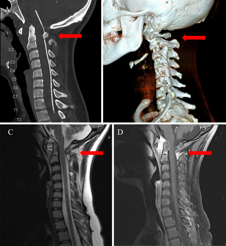
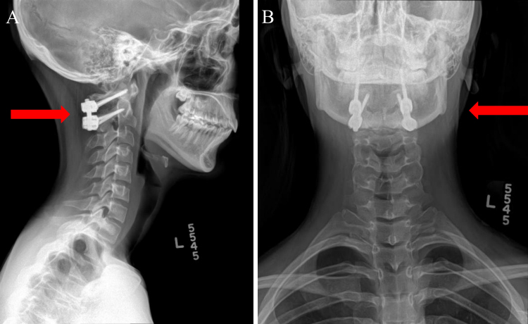
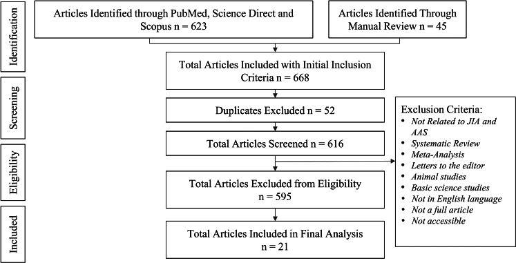
Observations: A 13-year-old female presented with progressive neck pain. Imaging revealed a 10-mm atlantodental interval on CT, along with hyperintensity and stretching of the transverse atlantal ligament on MRI. She underwent a posterior C1-2 open reduction and fusion. Subsequent rheumatological workup confirmed enthesitis-related JIA, based on polyarticular arthritis, HLA-B27 positivity, and elevated inflammatory markers. To contextualize this case, the authors performed a systematic review and meta-analysis of JIA-related AAS across 21 studies. The pooled incidence of AAS was 14%, with a mean age at JIA onset of 8.47 years and a female predominance of 62%. Enthesitis-related arthritis emerged as the most frequently reported subtype, and 94.4% of patients with AAS improved posttreatment.
Lessons: This case and supporting literature underscore the importance of early detection and multidisciplinary management of AAS in pediatric patients with JIA. Timely neurosurgical stabilization, combined with optimized immunosuppressive therapy, can prevent neurological compromise. Future research should focus on standardized diagnostic thresholds and outcome measures to guide best practices. https://thejns.org/doi/10.3171/CASE25121.
Keywords: atlantoaxial subluxation; cervical spine fusion; enthesitis-related arthritis; juvenile idiopathic arthritis; pediatric spine instability.
Figures
Similar articles
-
Biomechanical rationale for the pathology of rheumatoid arthritis in the craniovertebral junction.Spine (Phila Pa 1976). 2000 Jul 1;25(13):1607-16. doi: 10.1097/00007632-200007010-00003. Spine (Phila Pa 1976). 2000. PMID: 10870135
-
Atlantoaxial subluxation as an early manifestation in an adolescent with undifferentiated spondyloarthritis: a case report and review of the literature.J Med Case Rep. 2011 Jul 3;5:275. doi: 10.1186/1752-1947-5-275. J Med Case Rep. 2011. PMID: 21722401 Free PMC article.
-
Conservative management of idiopathic anterior atlantoaxial subluxation without neurological deficits in an 83-year-old female: A case report.J Can Chiropr Assoc. 2014 Mar;58(1):76-84. J Can Chiropr Assoc. 2014. PMID: 24587500 Free PMC article.
-
C1-C2 subluxation in enthesitis-related arthritis: two case reports and literature review of ten cases.Pediatr Rheumatol Online J. 2023 Aug 3;21(1):77. doi: 10.1186/s12969-023-00862-3. Pediatr Rheumatol Online J. 2023. PMID: 37537687 Free PMC article. Review.
-
Group A Streptococcus dysgalactiae subspecies equisimilis vertebral osteomyelitis accompanied by progressive atlantoaxial subluxation: A case report and literature review.Medicine (Baltimore). 2023 Aug 25;102(34):e34968. doi: 10.1097/MD.0000000000034968. Medicine (Baltimore). 2023. PMID: 37653834 Free PMC article. Review.
References
-
- Kolen ER Schmidt MH.. Rheumatoid arthritis of the cervical spine. Semin Neurol. 2002;22(2):179-186. - PubMed
-
- Neva MH Kaarela K Kauppi M.. Prevalence of radiological changes in the cervical spine—a cross sectional study after 20 years from presentation of rheumatoid arthritis. J Rheumatol. 2000;27(1):90-93. - PubMed
-
- Riew KD Hilibrand AS Palumbo MA Sethi N Bohlman HH.. Diagnosing basilar invagination in the rheumatoid patient. The reliability of radiographic criteria. J Bone Joint Surg Am. 2001;83(2):194-200. - PubMed
-
- Narváez JA, Narváez J, Serrallonga M.Cervical spine involvement in rheumatoid arthritis: correlation between neurological manifestations and magnetic resonance imaging findings. Rheumatology (Oxford). 2008;47(12):1814-1819. - PubMed
LinkOut - more resources
Full Text Sources
Research Materials
