. 2025 Jun;40(3):349-360.
doi: 10.1016/j.virs.2025.05.001. Epub 2025 May 17.

Characterization of a SARS-CoV-2 infection model in golden hamsters with diabetes mellitus

Affiliations

Characterization of a SARS-CoV-2 infection model in golden hamsters with diabetes mellitus

Hao-Feng Lin et al. Virol Sin. 2025 Jun.

Abstract

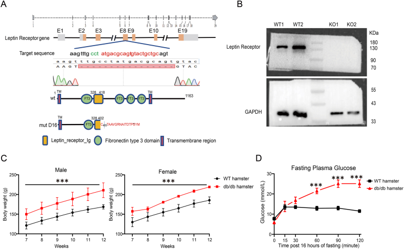
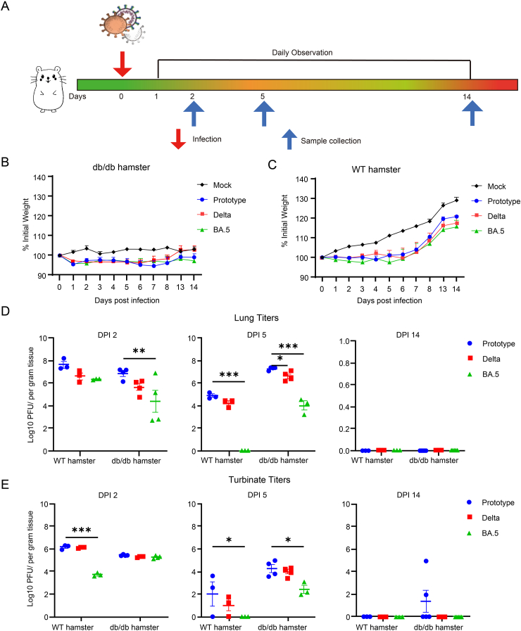
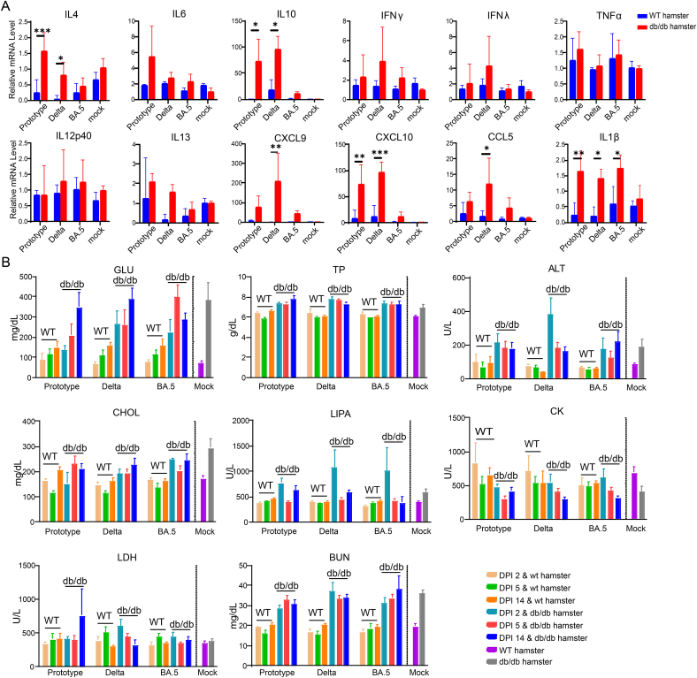
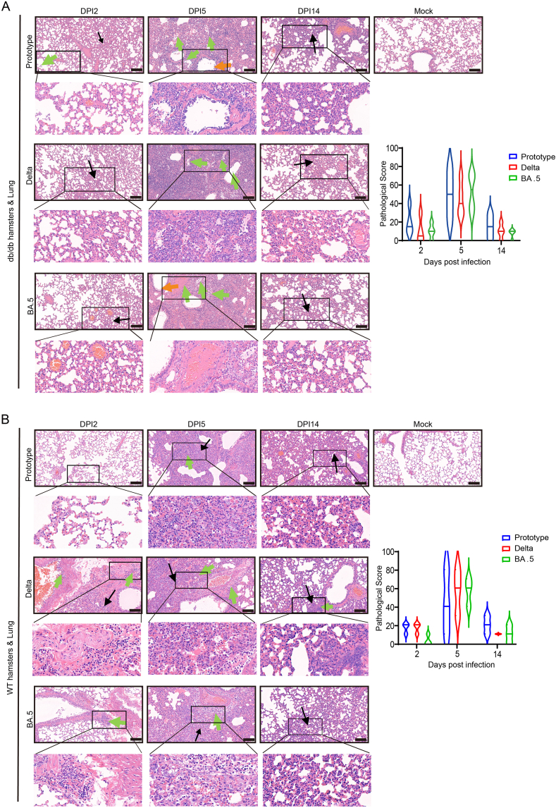
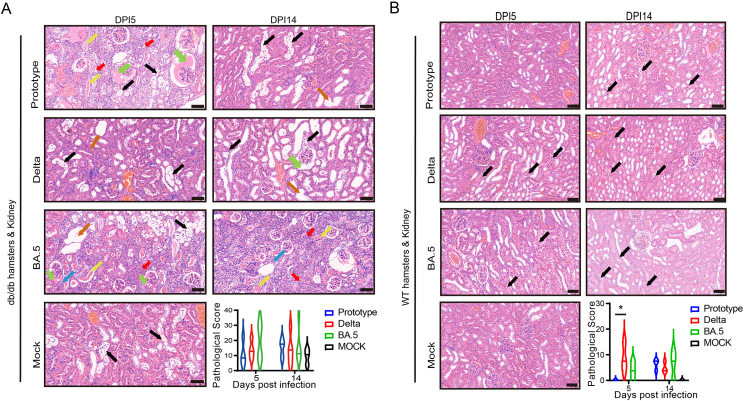
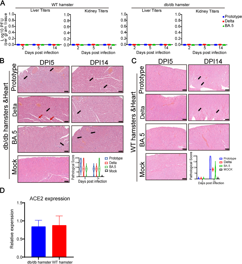
Being widespread across the globe, severe acute respiratory syndrome coronavirus 2 (SARS-CoV-2) keeps evolving and generating new variants and continuously poses threat to public health, especially to the population with chronic comorbidities. Diabetes mellitus is one of high-risk factors for severe outcome of coronavirus disease 2019 (COVID-19). Establishment of animal models that parallel the clinical and pathological features of COVID-19 complicated with diabetes is thus highly essential. Here, in this study, we constructed leptin receptor gene knockout hamsters with the phenotype of diabetes mellitus (db/db), and revealed that the diabetic hamsters were more susceptible to SARS-CoV-2 and its variants than wild-type hamsters. SARS-CoV-2 and its variants induced a stronger immune cytokine response in the lungs of diabetic hamsters than in wild-type hamsters. Comparative histopathology analyses also showed that infection of SARS-CoV-2 and the variants caused more severe lung tissue injury in diabetic hamsters, and may induce serious complications such as diabetic kidney disease and cardiac lesions. Our findings demonstrated that despite the decreased respiratory pathogenicity, the SARS-CoV-2 variants were still capable of impairing other organs such as kidney and heart in diabetic hamsters, suggesting that the risk of evolving SARS-CoV-2 variants to diabetic patients should never be neglected. This hamster model may help better understand the pathogenesis mechanism of severe COVID-19 in patients with diabetes. It will also aid in development and testing of effective therapeutics and prophylactic treatments against SARS-CoV-2 variants among these high-risk populations.

Keywords: Diabetes; Hamster; Multiorgan injury; Pneumonia; Severe acute respiratory syndrome coronavirus 2 (SARS-CoV-2).


Conflict of interest statement

Conflict of interest All authors declare that there are no competing interests.

Figures

Fig. 1
Fig. 1
Strategy for production of the leptin receptor-deficient golden hamsters by CRISPR/Cas9. A Structure of the golden hamster leptin receptor gene and target locus for sgRNA. Sequencing result showed 16 nucleotides deletion (mut16) in the leptin receptor-deficient (db/db) golden hamster lines, which resulted in leptin receptor protein mistranslation. B Western blots showed the loss of leptin receptor expression in subcutaneous inguinal adipose tissue of db/db hamsters. GAPDH was used as the loading control. C Under normal feeding conditions, including feeding with commercial common hamster food, identical rearing environments and feeding frequencies, the body weights of 7–12-week-old db/db hamsters (n ​= ​5) and WT hamsters (n ​= ​5) were routinely monitored. D Blood samples were collected from db/db hamsters (n ​= ​5, 2 males, 3 females) and WT hamsters (n ​= ​5, 2 males, 3 females) at various time points following a 16-h fasting period to monitor glucose levels. Error bars indicated the standard error. Statistical significance was measured by two-way ANOVA with multi-comparison model. ∗∗∗ P < 0.001.
Fig. 2
Fig. 2
db/db hamsters are highly susceptible to SARS-CoV-2 variants. Hamsters were divided into two groups, one for monitoring weight change and the survival rate, and the other for tissue sampling at various time points after infection. In the tissue sampling group, six-to-ten-week-old male and female db/db hamsters or wild-type (WT) hamsters were mock-infected or intranasally infected with 1 ​× ​105 TCID50 of SARS-CoV-2 prototype, SARS-CoV-2 Delta variant, or SARS-CoV-2 Omicron BA.5 variant, and the infected hamsters were sacrificed for tissue collection at 2, 5, and 14 DPI. A Scheme of animal infection. B, C For the survival monitoring, body weight and survival of db/db hamsters and wild-type hamsters were monitored daily until 14 DPI after challenge with either DMEM (Mock, 1 male and 2 females) or different SARS-CoV-2 strains (2 males and 3 females). D, E For tissue collection of the lung and turbinate at 2, 5, and 14 DPI, virus-infected db/db hamsters (6 males and 7 females for each strain) and WT hamsters (5 males and 4 females for each strain) were euthanized, with both male and female animals included at each sampling time point. Viral load in lung and turbinate of the infected WT and db/db hamsters were detected by plaque formation assay. Error bars indicate the standard error. Statistical significance was measured by two-way ANOVA with multi-comparison model. ∗P ​< ​0.05, ∗∗P ​< ​0.01, ∗∗∗P ​< ​0.001.
Fig. 3
Fig. 3
Testing of clinical blood biochemical indices and immune response in WT and db/db hamsters infected with SARS-CoV-2 variants. Six-to-ten-week-old male and female WT hamsters and db/db hamsters infected with 1 ​× ​105 TCID50 of SARS-CoV-2 prototype, SARS-CoV-2 Delta strain, or SARS-CoV-2 Omicron BA.5 strains, and then they were sacrificed for blood and lung tissue collection at 2, 5, and 14 DPI. A Cytokine and chemokine from infected-hamsters lung lysates at 5 DPI was quantified by qPCR. B Serum samples from infected hamsters at different time points were tested for some clinical blood biochemical indices. Error bars indicate the standard error. Statistical significance was measured by two-way ANOVA with multi-comparison model. ∗P ​< ​0.05, ∗∗P ​< ​0.01, ∗∗∗P ​< ​0.001.
Fig. 4
Fig. 4
The lung histopathological changes and pathological scoring in db/db hamsters and WT hamsters. A, B Six-to-ten-week-old male and female hamsters were mock-infected or intranasally infected with 1 ​× ​105 TCID50 of SARS-CoV-2 prototype, SARS-CoV-2 Delta variant, or SARS-CoV-2 Omicron BA.5 variant. Infected db/db hamsters (A) and WT hamsters (B) were sacrificed ​at 2, 5, and 14 DPI to examine the pathological changes in the lungs, while mock-infected hamsters were sacrificed at 14 DPI as the negative control group. Meanwhile, semiquantitative analysis and pathology scoring of the H&E-stained tissues section were performed. Images were collected using a Pannoramic MIDI system. In lung histopathology, black arrows indicate alveolar wall thickening and widening of the alveolar spacing; green arrows signify lymphocytic and granulocytic infiltration, and orange arrows denote bronchial epithelial cell necrosis. The scores were determined based on the percentage of inflammation in the pulmonary section by using the following 0–70 point scoring system: 0, no inflammation; 15, affected area (≤1 ​%); 30, affected area (>1 ​%, ≤10 ​%); 45, affected area (>10 ​%, ≤50 ​%); 60, affected area (>50 ​%). Additional 10 points were added when pulmonary edema, inflammatory cell infiltration, fibroplasia, and/or hyaline membrane formation were observed. The scale bar is 50 μm. Error bars indicate the standard error. Statistical significance was measured by two-way ANOVA. No significance was observed among different groups.
Fig. 5
Fig. 5
The kidney histopathological changes and pathological scoring in db/db hamsters and WT hamsters. A, B Six-to-ten-week-old male and female hamsters were mock-infected or intranasally infected with 1 ​× ​105 TCID50 of SARS-CoV-2 prototype, SARS-CoV-2 Delta variant, or SARS-CoV-2 Omicron BA.5 variant. Infected db/db hamsters (A) and WT hamsters (B) were sacrificed at 2, 5, and 14 DPI to examine the pathological changes in the kidneys, while mock-infected hamsters were sacrificed at 14 DPI as the negative control group. Meanwhile, semiquantitative analysis and pathology scoring of the H&E-stained tissues section were conducted. Images were collected using a Pannoramic MIDI system. In kidney histopathology, black arrows represent necrosis of renal tubular epithelial cells and cytoplasmic vacuolization; green arrows indicate dilatation of the glomerular capsule filled with eosinophilic secretions; red arrows signify atrophy of cortical medullary tubules; yellow arrows indicate the formation of a proteinaceous tubular pattern; orange arrows denote tubule dilatation with irregular lumens and flattening of tubular epithelial cells, and blue arrows indicate lymphocyte infiltration. The scale bar is 50 μm. Error bars indicate the standard error. Statistical significance was measured by two-way ANOVA. ​∗ P < 0.05.
Supplementary Fig. S1
Other tissue pathologic changes and viral titers in infected db/db hamsters and WT hamsters. A Six-to-ten-week-old male and female hamsters were mock-infected or intranasally infected with 1 ​× ​105 TCID50 of SARS-CoV-2 prototype, SARS-CoV-2 Delta variant, or SARS-CoV-2 Omicron BA.5 variant. The virus-infected hamsters were sacrificed for tissue collection (liver and kidney) at 2, 5, and 14 DPI, respectively. Viral load in liver and kidney of the infected WT and db/db hamsters were detected by plaque formation assay. B, C db/db hamsters (B) and WT hamsters (C) were sacrificed to examine the pathological changes in the hearts at 2, 5, and 14 DPI. Meanwhile, semiquantitative analysis and pathology scoring of the H&E-stained tissues section were conducted. Images were collected using a Pannoramic MIDI system. In heart histopathology, black arrows signify hydropic degeneration of cardiomyocytes, while red arrows denote breaks in myocardial fibers. The scale bar is 50 μm. D The nucleic acids were extracted from WT and db/db hamsters (n ​= ​8) lung tissues, followed by RT-qPCR analysis, and the relative gene expression level of ACE2 was quantified. Error bars indicate the standard error. Statistical significance was measured by two-way ANOVA with multi-comparison model. No significance was observed among different groups.

Similar articles

References

    1. Allard R., Leclerc P., Tremblay C., Tannenbaum T.N. Diabetes and the severity of pandemic influenza A (H1N1) infection. Diabetes Care. 2010;33:1491–1493. - PMC - PubMed
    1. Alraddadi B.M., Watson J.T., Almarashi A., Abedi G.R., Turkistani A., Sadran M., Housa A., Almazroa M.A., Alraihan N., Banjar A., Albalawi E., Alhindi H., Choudhry A.J., Meiman J.G., Paczkowski M., Curns A., Mounts A., Feikin D.R., Marano N., Swerdlow D.L., Gerber S.I., Hajjeh R., Madani T.A. Risk factors for primary Middle East respiratory syndrome coronavirus illness in humans, Saudi arabia, 2014. Emerg. Infect. Dis. 2016;22:49–55. - PMC - PubMed
    1. Barron E., Bakhai C., Kar P., Weaver A., Bradley D., Ismail H., Knighton P., Holman N., Khunti K., Sattar N., Wareham N.J., Young B., Valabhji J. Associations of type 1 and type 2 diabetes with COVID-19-related mortality in England: a whole-population study. Lancet Diabetes Endocrinol. 2020;8:813–822. - PMC - PubMed
    1. Buetti N., Trimboli P., Mazzuchelli T., Lo Priore E., Balmelli C., Trkola A., Conti M., Martinetti G., Elzi L., Ceschi A., Consonni V., Ogna A., Forni-Ogna V., Bernasconi E. Diabetes mellitus is a risk factor for prolonged SARS-CoV-2 viral shedding in lower respiratory tract samples of critically ill patients. Endocrine. 2020;70:454–460. - PMC - PubMed
    1. Buetti N., Wicky P.H., Le Hingrat Q., Ruckly S., Mazzuchelli T., Loiodice A., Trimboli P., Forni Ogna V., De Montmollin E., Bernasconi E., Visseaux B., Timsit J.F. SARS-CoV-2 detection in the lower respiratory tract of invasively ventilated ARDS patients. Crit. Care. 2020;24:610. - PMC - PubMed

Supplementary concepts

LinkOut - more resources