. 2025 May 19;15(1):17286.
doi: 10.1038/s41598-025-00707-x.

NAT10 promotes hepatocellular carcinoma progression by modulating the ac4C-DDIAS-PI3K-Akt axis

Affiliations

NAT10 promotes hepatocellular carcinoma progression by modulating the ac4C-DDIAS-PI3K-Akt axis

Yue Tao et al. Sci Rep. .

Abstract

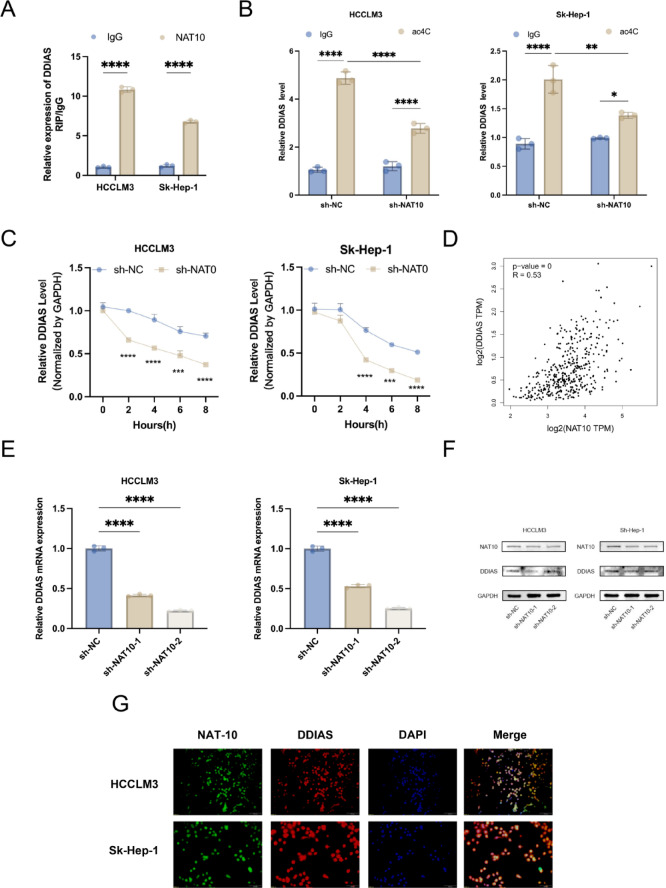
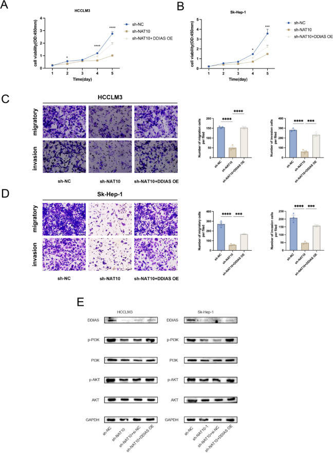
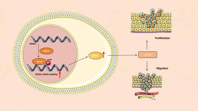
Primary liver cancer (PLC) is a prevalent tumor globally, ranking third in cancer-related mortality. The role of N4-acetylcysteine (ac4C) and N-acetyltransferase 10 (NAT10) in hepatocellular carcinoma (HCC) progression, migration, and invasion requires further elucidation. High NAT10 expression correlated with poor prognosis in HCC patients. Knockdown of NAT10 hindered HCC cell proliferation. AcRIP-seq screening revealed DDIAS as a significant downstream target of NAT10. Decreased NAT10 levels reduced DDIAS mRNA stability, leading to decreased proliferation, migration, and invasion of HCC cells upon DDIAS knockdown. Ectopic expression of DDIAS counteracted the effects of NAT10 knockdown by modulating the PI3K/AKT pathway. NAT10 was found to be elevated in HCC tissues compared to normal tissues, promoting HCC progression and correlating with shorter overall survival in patients. Mechanistically, NAT10 regulated HCC progression through the ac4C-DDIAS-PI3K-AKT axis.

Keywords: Acetylation; DDIAS; Hepatocellular carcinoma; NAT10; PI3K-AKT; ac4C.


Conflict of interest statement

Declarations. Competing interests: The authors declare no competing interests.

Figures

Fig. 1
Fig. 1
NAT10 was highly expressed and prognostic values in HCC. (A) The web-based tool GEPIA was utilized to investigate NAT10 mRNA expression levels in HCC tissues. (B) The Kaplan-Meier plotter, an online resource, was used to determine the relationship between NAT10 mRNA expression and overall survival (left) and progression-free survival (right) in HCC patients. (C) Kaplan-Meier survival analysis was conducted based on NAT10 expression in early-stage HCC patients (from TCGA cohort, stages 1 and 2). (D) Western blot analysis was performed on six HCC sample pairs to assess NAT10 expression. (E) Representative images depicting NAT10 IHC staining in HCC samples were presented, with scale bars measuring 100 μm. (F) IHC scores for matched HCC and normal tissues (n = 77) were calculated based on NAT10 staining. (G) Kaplan-Meier analysis of HCC patient progression-free survival relative to NAT10 expression levels was carried out (n = 77). The median of IHC scores served as the cutoff for group classification. Knockdown of NAT10 was observed to inhibit proliferation, migration, and invasion capacities in HCC cells during in vitro studies. (A, B) RT-qPCR and Western blot assays confirmed NAT10 expression levels in shNAT10 HCC cells. (C) The CCK8 assay assessed the impact of NAT10 knockdown on the proliferation of HCC cells. (D, E) Transwell assays evaluated the effect of NAT10 knockdown on the migratory (D) and invasive (E) abilities of HCC cells, with a scale bar of 200 μm.
Fig. 2
Fig. 2
Knockdown of NAT10 hindered the proliferation, migration, and invasion capabilities of HCC cells in vitro. (A, B) RT-qPCR and Western blotting assays were used to verify NAT10 expression in shNAT10 HCC cells. (C) The CCK8 assay was employed to explore the influence of NAT10 knockdown on the proliferative capacity of HCC cells. (D, E) Transwell assays were conducted to scrutinize the impact of NAT10 knockdown on HCC cell migratory (D) and invasive (E) capacities. Scale bar: 200 μm.
Fig. 3
Fig. 3
ac4C-seq and RNA-seq analysis demonstrated NAT10 as a potential target of DDIAS. (A) RNA-sequencing was utilized to examine the differential gene expression in HCCLM3 cells transfected with either NC or NAT10 shRNA. (B) The ac4C peaks were predominantly found within the CDS and 30UTR regions of HCC cells. (C) The proportion of mRNAs containing varying quantities of ac4C peaks was determined. (D) A Venn diagram was utilized to identify the mRNAs exhibiting alterations in both ac4C peak levels and gene expression. (E) The star plot displayed the distribution of genes showing differential ac4C peaks (hyper or hypo; Y-axis; fold change > 1.5 or 2 or < 0.5, P < 0.05) in the NAT10-downexpressing group compared to the control group. (F) The location of the ac4C peak on DDIAS was analyzed using IGV.
Fig. 4
Fig. 4
NAT10 controlled DDIAS expression in an acetylation-dependent manner. (A) Binding capacity between DDIAS mRNA and NAT10 in HCC cells was determined using RIP assay. (B) acRIP-qPCR was used to analyze the impact of NAT10 knockdown on acetylation levels of DDIAS in HCC cells. (C) qPCR was performed to examine the effect of NAT10 knockdown on DDIAS half-life in HCC cells. (D) The relationship between DDIAS and NAT10 expression in The Cancer Genome Atlas database for HCC was investigated. (E) qPCR was utilized to analyze the changes in DDIAS mRNA expression upon NAT10 knockdown. (F) DDIAS protein levels were measured post-NAT10 knockdown. Data was presented as means ± SD. (G) Immunofluorescence assay showed co-localization of NAT10 (green) and DDIAS (red) with DAPI (blue) staining marking the nucleus (scalebar = 100 μm).
Fig. 5
Fig. 5
DDIAS facilitated both the proliferation and invasion of HCC. (A) DDIAS mRNA levels were evaluated in HCC tissues using the GEPIA online tool. (B) The potential correlation between DDIAS mRNA expression and overall survival (left) as well as progression-free survival (right) in HCC was examined via the Kaplan-Meier plotter online tool. (C, D) qPCR (C) and western blot assays (D) were utilized to measure DDIAS expression in HCC cells and Lx2 cells. (E) Representative images displaying DDIAS IHC staining in HCC samples were presented (scale bars: 100 μm).
Fig. 6
Fig. 6
By influencing the DDIAS-PI3K-AKT pathway, NAT10 controlled the invasion and proliferation of HCC.(A, B) Using the CCK8 assay, the impact of DDIAS knockdown on the ability of HCC cells to proliferate was investigated (C, D) Using a transwell experiment, the effect of DDIAS knockdown on the migratory (C) and invasive (D) potential of HCC cells was investigated. Scale bar: 200 μm. (E, F) Activation of PI3K/Akt signaling pathway as detected after (E) DDIAS knockdown or (F) NAT10 knockdown in HCCLM3 and Sk-Hep-1 cells.
Fig. 7
Fig. 7
The schematic below depicts the role NAT10 plays in modulating PI3K/Akt signaling in HCC progression. (A, B) CCK8 assay was used to examine the reversal effect of DDIAS overexpression on proliferative capacity of shNAT10 HCC cells.(C, D) Transwell assays were used to determine the reversal effects of DDIAS overexpression on the migratory (C) and invasive (D) capacities of shNAT10 cells. Scale bar: 100 μm (E) Western blot analysis was conducted to assess the impact of DDIAS overexpression on the protein levels of key Akt pathway components in hepatocellular carcinoma cells.
Fig. 8
Fig. 8
A diagram demonstrates that NAT10 has the potential to be a valuable focus for enhancing HCC treatment by acetylating and stabilizing DDIAS mRNA to boost its levels and regulate the PI3K-AKT pathway.This promotes the progression and proliferation of HCC.

References

    1. Sung, H. et al. Global Cancer statistics 2020: GLOBOCAN estimates of incidence and mortality worldwide for 36 cancers in 185 countries. CA Cancer J. Clin.71, 209–249 (2021). - PubMed
    1. Petrick, J. L. et al. International trends in hepatocellular carcinoma incidence, 1978–2012. Int. J. Cancer. 147, 317–330 (2020). - PMC - PubMed
    1. Llovet, J. M. et al. Hepatocellular carcinoma. Nat. Rev. Dis. Primers. 7, 6 (2021). - PubMed
    1. Roderburg, C., Özdirik, B., Wree, A., Demir, M. & Tacke, F. Systemic treatment of hepatocellular carcinoma: from Sorafenib to combination therapies. Hepat. Oncol.7 (2), HEP20 (2020). - PMC - PubMed
    1. Xie, D. Y., Ren, Z. G., Zhou, J., Fan, J. & Gao, Q. 2019 Chinese clinical guidelines for the management of hepatocellular carcinoma: updates and insights. Hepatobiliary Surg Nutr.9, 452–463 (2020). - PMC - PubMed

MeSH terms

Substances

LinkOut - more resources